Sporothrix davidellisii : A new pathogenic species belonging to the Sporothrix pallida complex
- PMID: 40216404
- PMCID: PMC12015470
- DOI: 10.1093/mmy/myaf034
Sporothrix davidellisii : A new pathogenic species belonging to the Sporothrix pallida complex
Abstract
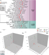
Sporothrix species (Ascomycota, Ophiostomatales) are dimorphic fungi with diverse ecological niches, ranging from mammalian, plant, and insect pathogens to fungicolous organisms. Here, we describe Sporothrix davidellisii (CBS 147636T), a novel pathogenic species within the S. pallida complex isolated from a case of feline sporotrichosis in Melbourne, Australia. Phylogenetic analyses based on ITS, β-tubulin (BT2), calmodulin (CAL), and translation elongation factor 1-α (EF1-α) sequences confirmed its distinctiveness, with ITS sequence identity to its closest relative (S. chilensis) not exceeding 97.6%. The assembled genome is 39.02 Mb (eight contigs) with a 27.2 kb mitochondrial genome and a total of 12,631 predicted genes. Genetic diversity analyses revealed moderate nucleotide variation in the ITS region (π = 0.055), greater diversity in BT2 (π = 0.098), and CAL (π = 0.118), supporting its status as a unique species. Morphological studies revealed distinctive characteristics differentiating S. davidellisii from its nearest relatives, including elongated clavate sympodial conidia and sessile conidia. Notably, S. davidellisii exhibits yeast-like growth at 37°C, forming ellipsoid to ovoid budding cells in liquid media, although cigar-shaped yeasts, characteristic of highly virulent Sporothrix species, are rarely observed. This ability to transition to a yeast-like form, combined with its high-temperature tolerance (growth up to 40°C), underscores its opportunistic pathogenic potential. The pathogenic role of S. davidellisii highlights the importance of monitoring atypical Sporothrix infections in feline hosts, which may serve as environmental sentinels for emerging fungal pathogens. These findings expand the taxonomy of Sporothrix, contributing to our understanding of the evolutionary complexity and zoonotic potential of species within the S. pallida complex.
Keywords: Sporothrix davidellisii; Sporothrix pallida complex; dimorphic fungi Ophiostomatales; feline sporotrichosis; sporotrichosis.
Plain language summary
The ascomycetous genus Sporothrix comprises dimorphic fungi with diverse ecological niches, ranging from mammalian, plant, and insect pathogens to fungicolous organisms. We describe Sporothrix davidellisii, a pathogenic member of the S. pallida complex isolated from a case of feline sporotrichosis.
© The Author(s) 2025. Published by Oxford University Press on behalf of The International Society for Human and Animal Mycology.
Conflict of interest statement
None of the authors declare a conflict of interest related to the topic described in this publication.
Figures


Similar articles
-
Phylogenetic analysis reveals a high prevalence of Sporothrix brasiliensis in feline sporotrichosis outbreaks.PLoS Negl Trop Dis. 2013 Jun 20;7(6):e2281. doi: 10.1371/journal.pntd.0002281. Print 2013. PLoS Negl Trop Dis. 2013. PMID: 23818999 Free PMC article.
-
Sporothrix chilensis sp. nov. (Ascomycota: Ophiostomatales), a soil-borne agent of human sporotrichosis with mild-pathogenic potential to mammals.Fungal Biol. 2016 Feb;120(2):246-64. doi: 10.1016/j.funbio.2015.05.006. Epub 2015 Jun 11. Fungal Biol. 2016. PMID: 26781380
-
Comparative genomics of the major fungal agents of human and animal Sporotrichosis: Sporothrix schenckii and Sporothrix brasiliensis.BMC Genomics. 2014 Oct 29;15:943. doi: 10.1186/1471-2164-15-943. BMC Genomics. 2014. PMID: 25351875 Free PMC article.
-
Molecular identification of the Sporothrix schenckii complex.Rev Iberoam Micol. 2014 Jan-Mar;31(1):2-6. doi: 10.1016/j.riam.2013.09.008. Epub 2013 Nov 19. Rev Iberoam Micol. 2014. PMID: 24270070 Review.
-
Origin and distribution of Sporothrix globosa causing sapronoses in Asia.J Med Microbiol. 2017 May;66(5):560-569. doi: 10.1099/jmm.0.000451. Epub 2017 May 22. J Med Microbiol. 2017. PMID: 28327256 Review.
References
-
- Machado TC, Gonçalves SS, de Carvalho JA, et al. Insights from cutting-edge diagnostics and epidemiology of sporotrichosis and taxonomic shifts in Sporothrix. Curr Fung Infect Rep. 2025; 19: 1–23.
MeSH terms
Substances
Grants and funding
- FAPESP 2017/27265-5/São Paulo Research Foundation
- CNPq 433276/2018-5/National Council for Scientific and Technological Development
- CNPq 405934/2022-0/National Institute of Science and Technology in Human Pathogenic Fungi, Brazil
- CAPES 88887.159096/2017-00/Coordination for the Improvement of Higher Education Personnel
- CNPq 314089/2023-3/CNPq
LinkOut - more resources
Full Text Sources
Research Materials
Miscellaneous

