. 2025 Jun 16;31(7):923-932.
doi: 10.1261/rna.080442.125.

The m6A-binding protein YTHDF3 modulates the cardiac response to stress

Affiliations

The m6A-binding protein YTHDF3 modulates the cardiac response to stress

Charles P Rabolli et al. RNA. .

Abstract

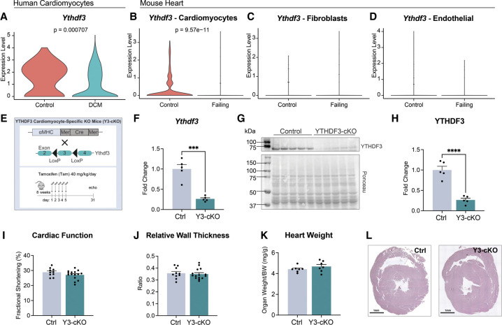
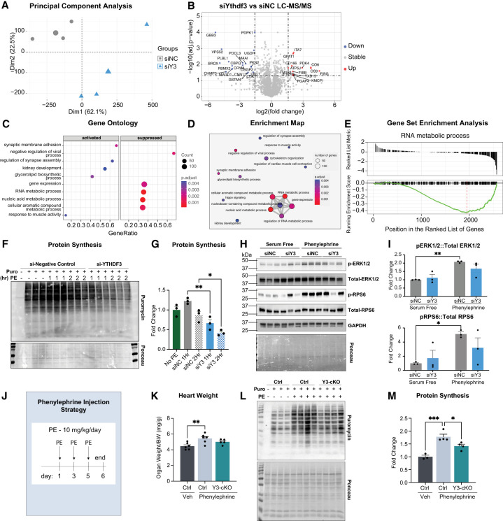
Transcriptional regulation of gene expression has long been studied; however, only recently has the impact of chemical mRNA modification on protein synthesis emerged. Among posttranscriptional modifications, methylation of the N6-adenosine site of mRNA (m6A) is very prevalent in eukaryotes and plays a critical role in the heart. To date, the mechanism through which m6A controls cardiac function remains elusive. The fate of m6A-modified mRNAs is regulated by members of the YTH domain family (YTHDF), such as YTHDF3. Here we report that mice with a cardiomyocyte-specific deletion of YTHDF3 have attenuated pathological remodeling following pressure overload injury. Mechanistically, we found that YTHDF3 regulates global stress-induced protein synthesis, and that this protein controls cardiomyocyte size. Altogether, this study uncovered a potential cardioprotective role for YTHDF3 inhibition and improves our understanding on the mechanism through which m6A impacts cardiac function.

Keywords: METTL3; YTHDF3; cardiac; heart; m6A.


Figures

FIGURE 1.
FIGURE 1.
(A) Single cardiomyocyte RNA expression for Ythdf3 from human nonfailing (control) and failing (DCM, dilated cardiomyopathy) hearts. (B–D) Single-cell RNA expression for Ythdf3 from murine nonfailing (control) and failing hearts, following transverse aortic constriction. (E) Schematic of cardiomyocyte-specific Ythdf3 knockout mouse model (Y3-cKO). (F) Confirmation of Ythdf3 knockout at the mRNA level using qPCR. Rpl7 was used as a housekeeping gene. n = 5 per group. (G,H) Western blot and quantification of YTHDF3 protein knockout. Ponceau was used as a loading control. n = 5 per group. (I,J) Echocardiography analysis of cardiac function by fractional shortening or relative wall thickness after 1 month of deletion. n = (10; 15) for (Ctrl; Y3-cKO). (K) Postmortem gravimetric analysis of heart weights 1 month after gene deletion, normalized to body weight (BW). n = 7 per group. (L) Hematoxylin and eosin staining 1 month after Ythdf3 knockout. Data shown as mean ± SEM. Unpaired t-test (AD,F,HK) was used, (*) P < 0.05, (**) P < 0.01, (***) P < 0.001, (****) P < 0.0001.
FIGURE 2.
FIGURE 2.
(A–C) Echocardiography analysis of cardiac function by fractional shortening and relative wall thickness following pressure overload injury by transverse aortic constriction (TAC). n = (7; 5; 8; 8) for (Ctrl Sham; Y3-cKO Sham; Ctrl TAC; Y3-cKO TAC). (D,E) Postmortem gravimetric analysis of heart and lung weights following TAC, normalized to tibia length (TL). n = (7; 5; 7; 8) for (Ctrl Sham; Y3-cKO Sham; Ctrl TAC; Y3-cKO TAC). (F,G) Wheat Germ Agglutinin staining and quantification following sham or TAC surgeries. n = (4; 5; 9; 5) for (Ctrl Sham; Y3-cKO Sham; Ctrl TAC; Y3-cKO TAC). (H,I) Picrosirius red staining and quantification following sham or TAC surgeries. n = (4; 5; 7; 8) for (Ctrl Sham; Y3-cKO Sham; Ctrl TAC; Y3-cKO TAC). (J–O) qPCR of the fibrotic markers Col1a1, Postn, and Ctgf and the inflammatory markers IL-6, Cxcl10, and IL-1β. Rpl7 was used as a housekeeping gene. n = (7; 5; 9; 9) for (Ctrl Sham; Y3-cKO Sham; Ctrl TAC; Y3-cKO TAC). Data shown as mean ± SEM. Two-way ANOVA with multiple comparisons test was used (A,CE,G,IO), (*) P < 0.05, (**) P < 0.01, (***) P < 0.001, (****) P < 0.0001.
FIGURE 3.
FIGURE 3.
(A) Design of neonatal rat ventricular cardiomyocytes experiments. (siNC) siRNA noncoding control, siYthdf3 (siY3) siRNA targeting Ythdf3. Figure made in Biorender. (B,C) Western blot confirming knockdown of YTHDF3 at the protein level, following siRNA treatment. n = 2 per group per condition. SDHB is used as a loading control. (D,E) Immunofluorescence images and quantification of a cross-sectional area following designated treatments. Minimum of 50 cells quantified per group per condition. (F,G) qPCR quantification of the designated genes in the assigned conditions. Rpl7 was used as a housekeeping gene. n = 4 per group per condition. Data shown as mean ± SEM. Unpaired t-test was used (C,EH), (*) P < 0.05, (**) P < 0.01, (***) P < 0.001, (****) P < 0.0001.
FIGURE 4.
FIGURE 4.
(A) Principal component analysis highlights clustering of groups following control or Ythdf3 knockdown treatments. n = 3 per group. (B) Volcano plot of mass spectrometry results following 2% serum stimulation of neonatal rat ventricular myocytes. n = 3 per group. (C–E) Gene enrichment analyses highlights mRNA metabolism as a key pathway that is regulated by YTHDF3. Analysis performed using clusterProfiler package in R. (F,G) Puromycin incorporation assay to test protein synthesis. n = 3 per condition per group; n = 1 for no puromycin control. (H,I) Western blots and quantification for phospho- and total-ERK1/2, and RPS6, normalized to GAPDH on neonatal rat ventricular myocytes treated with control siRNA (siNC) or siRNA against YTHDF3 (siY3) in serum free with and without 1 h of phenylephrine stimulation. n = 3 per group. (J) Schematic of adult in vivo phenylephrine (PE) injection strategy. (K) Heart weight following PE injections, normalized to body weight (BW). n = (6; 6; 5) for (Ctrl vehicle; Ctrl PE; Y3-cKO PE). (L,M) Puromycin western blot and quantification of adult heart following PE injections. Ponceau was used as loading control. n = (1; 3; 4; 4) for (no puromycin Ctrl; Ctrl vehicle; Ctrl PE; Y3-cKO PE). Data shown as mean ± SEM. One-way ANOVA was used (G,K,M) and two-way ANOVA was used (I), (*) P < 0.05, (**) P < 0.01, (***) P < 0.001, (****) P < 0.0001.
Charles P. Rabolli
Charles P. Rabolli

References

    1. Aufgebauer CJ, Bland KM, Horner SM. 2024. Modifying the antiviral innate immune response by selective writing, erasing, and reading of m6A on viral and cellular RNA. Cell Chem Biol 31: 100–109. 10.1016/j.chembiol.2023.12.004 - DOI - PMC - PubMed
    1. Berulava T, Buchholz E, Elerdashvili V, Pena T, Islam MR, Lbik D, Mohamed BA, Renner A, von Lewinski D, Sacherer M, et al. 2020. Changes in m6A RNA methylation contribute to heart failure progression by modulating translation. Eur J Heart Fail 22: 54–66. 10.1002/ejhf.1672 - DOI - PubMed
    1. Cappannini A, Ray A, Purta E, Mukherjee S, Boccaletto P, Moafinejad SN, Lechner A, Barchet C, Klaholz BP, Stefaniak F, et al. 2024. MODOMICS: a database of RNA modifications and related information. 2023 update. Nucleic Acids Res 52: D239–D244. 10.1093/nar/gkad1083 - DOI - PMC - PubMed
    1. Chang G, Shi L, Ye Y, Shi H, Zeng L, Tiwary S, Huse JT, Huo L, Ma L, Ma Y, et al. 2020. YTHDF3 induces the translation of m6A-enriched gene transcripts to promote breast cancer brain metastasis. Cancer Cell 38: 857–871.e7. 10.1016/j.ccell.2020.10.004 - DOI - PMC - PubMed
    1. Chen L, Gao Y, Xu S, Yuan J, Wang M, Li T, Gong J. 2023. N6-methyladenosine reader YTHDF family in biological processes: structures, roles, and mechanisms. Front Immunol 14: 1162607. 10.3389/fimmu.2023.1162607 - DOI - PMC - PubMed

LinkOut - more resources