. 2025 Apr 11;14(1):9.
doi: 10.1038/s41389-025-00554-5.

Myeloma mesenchymal stem cells' bioenergetics afford a novel selective therapeutic target

Affiliations

Myeloma mesenchymal stem cells' bioenergetics afford a novel selective therapeutic target

Oded Komemi et al. Oncogenesis. .

Abstract

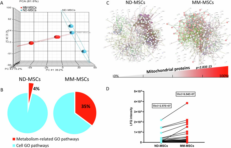
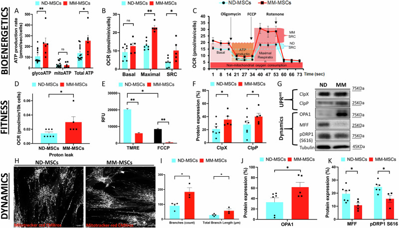
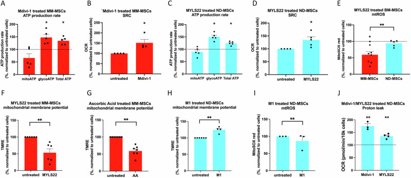
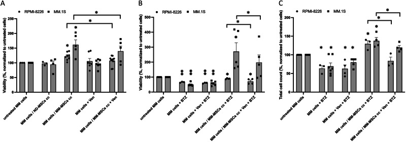
Bone-marrow mesenchymal stem cells (BM-MSCs) rely on glycolysis, yet their trafficked mitochondria benefit recipient cells' bioenergetics in regenerative and cancerous settings, most relevant to BM-resident multiple myeloma (MM) cells. Fission/fusion dynamics regulate mitochondria function. Proteomics demonstrates excessive mitochondrial processes in BM-MSCs from MM patients compared to normal donors (ND). Thus, we aimed to characterize BM-MSCs (ND, MM) mitochondrial fitness, bioenergetics and dynamics with a focus on therapeutics. MM-MSCs displayed compromised mitochondria evidenced by decreased mitochondrial membrane potential (ΔΨm) and elevated proton leak. This was accompanied by stimulation of stress-coping mechanisms: spare respiratory capacity (SRC), mitochondrial fusion and UPRmt. Interfering with BM-MSCs mitochondrial dynamics equilibrium demonstrated their significance to bioenergetics and fitness according to the source. While ND-MSCs depended on fission, reducing MM-MSCs fusion attenuated glycolysis, OXPHOS and mtROS. Interestingly, optimization of mtROS levels is central to ΔΨm preservation in MM-MSCs only. MM-MSCs also demonstrated STAT3 activation, which regulates their OXPHOS and SRC. Targeting MM-MSC' SRC with Venetoclax diminished their pro-MM support and sensitized co-cultured MM cells to Bortezomib. Overall, MM-MSCs distinct mitochondrial bioenergetics are integral to their robustness. Repurposing Venetoclax as anti-SRC treatment in combination with conventional anti-MM drugs presents a potential selective way to target MM-MSCs conferred drug resistance.


Conflict of interest statement

Competing interests: The authors declare no competing interests. Ethics approval and consent to participate: All procedures were performed in compliance with relevant laws and institutional guidelines and have been approved by the Meir Medical Center Helsinki Committee (REF: 0205-12-MMC ; REF: 0045-11-MMC). All patients or their guardians/legally authorized representatives/next of kin provided written informed consent for participation in the study and the use of samples.

Figures

Fig. 1
Fig. 1. Mass spectrometry analysis of MM-MSCs demonstrated increase in mitochondrial-related metabolic pathways and proteins.
A Principal component analysis (PCA) graph presents clear segregation of MM-MSCs and ND-MSCs mass spectrometry data according to source. B The BM-MSCs (ND and MM-MSCs) mass spectrometry data (differentially expressed proteins, p < 0.05 (FDR), N = 3 each) was assessed for cellular component and pathways using WebGestalt. Pie graphs present the percentage of metabolism-related GO pathways (red) out of all cell pathways (Cyan) in BM-MSCs. C STRING functional enrichment analysis of BM-MSCs upregulated proteins (FC ≥ 1.25, p < 0.05) included in the “Mitochondrion” cellular component STRING category (p = 2.16E−17, red nodes). D Median values of 63 protein levels (mass spectrometry) included in the BM-MSCs “Mitochondrion” cellular component category (WebGestalt, GO:0005739, p = 0) presented individually by shape and color.
Fig. 2
Fig. 2. MM-MSCs display increased bioenergetics, mitochondrial stress adaptation and elevated fusion.
ND (cyan) and MM (red) MSCs (n≧5) were subjected to (A) Assessment of cellular glycoATP, mitoATP and TotalATP production rates (Seahorse XF Real-Time ATP Rate Assay Kit). B Assessment of oxygen consumption rates (OCR) and determination of Basal respiration, Maximal respiration and SRC (Seahorse XF Cell Mito Stress Test Kit). C Representative graph of Seahorse XF Cell Mito Stress Test Kit. D Assessment of Proton leak (Seahorse XF Cell Mito Stress Test Kit). E Analysis of mitochondrial membrane potential (TMRE). F Quantification of UPRmt factors’ immunoblotting (ClpX and ClpP). G Representative Immunoblots of UPRmt factors (ClpX and ClpP) and Fusion/Fission regulators (OPA1, MFF and pDRP1S616). H Representative confocal images of BM-MSCs mitochondria (ND and MM, Mitotracker Red CMXros). I Analysis of mitochondria Branches and Total Branch Length (ND and MM confocal images, Imagej/FUJI Mitochondrial Analyzer tool). J Quantification of mitochondrial fusion regulator OPA1 levels (immunoblotting). K Quantification of mitochondrial fission regulators MFF and pDRP1S616 levels (immunoblotting). Immunoblots are cropped for visual ease. All immunoblotting results were normalized to tubulin and are expressed as percent (Mean ± SE, n ≥ 3) of protein levels in MM-MSCs compared to ND-MSCs. Asterisks depicted statistical significance *p < 0.05; **p < 0.01.
Fig. 3
Fig. 3. Intervention with fission/fusion balance differentially affects BM-MSCs bioenergetics, mtROS and ΔΨm.
ND (cyan) and MM (red) MSCs (n≧3) were treated with Mdivi-1 (mitochondrial fission inhibitor, 50 µM; 24 h), MYLS22 (mitochondrial fusion inhibitor, 50 µM; 24 h), M1 (mitochondrial fusion promoter, 5 µM; 24 h), Ascorbic Acid (AA) (ROS scavenger, 500 µM; 24 h) and analyzed for (A) MM-MSCs glycoATP, mitoATP and TotalATP production rates following Mdivi-1 treatment (Seahorse XF Real-Time ATP Rate Assay Kit) (B) MM-MSCs SRC following Mdivi-1 treatment (Seahorse XF Cell Mito Stress Test Kit). C ND-MSCs glycoATP, mitoATP and TotalATP production rates following MYLS22 treatment (Seahorse XF Real-Time ATP Rate Assay). D ND-MSCs SRC following MYLS22 treatment (Seahorse XF Cell Mito Stress Test Kit). E ND and MM-MSCs mtROS levels following MYLS22 treatment (mitoSOX red). F MM-MSCs mitochondrial membrane potential following MYLS22 treatment (TMRE). G MM-MSCs mitochondrial membrane potential following AA treatment (TMRE). H ND-MSCs mitochondrial membrane potential following M1 treatment (TMRE). I ND-MSCs mtROS levels following M1 treatment (mitoSOX red). J ND-MSCs proton leak following Mdivi-1/MYLS22 treatment (Seahorse XF Cell Mito Stress Test Kit). Results are expressed as percentage (Mean ± SE, n ≥ 3) and normalized to untreated cells represented by control bar (100%) or dashed line. Asterisks depicted statistical significance *p < 0.05; **p < 0.01.
Fig. 4
Fig. 4. Venetoclax mitigates MM-MSCs support of adjacent MM cells’ survival and sensitizes them to anti-MM drug Bortezomib.
ND and MM MSCs (n ≥ 3) were pre-treated with Venetoclax (Ven) (SRC inhibitor, 0.5 µM; 24 h) then co-cultured with RPMI-8226 or MM.1S MM cell lines (MM cells) in the presence of Ven for 48 h or added with Bortezomib (BTZ)(anti-MM drug, 5 nM; last) for the last 24 h of 72 h of co-culture. Untreated MM cells alone or in co-culture served as controls. A Ven’s effect on co-cultured (with ND/MM-MSCs, 72 h) MM cells’ Viability (Presto-Blue). B, C Ven ± BTZ effect on co-cultured (with MM-MSCs, 72 h) MM cells’ Viability (Presto-Blue) and total cell count (Trypan-Blue). Cell counts and viability were normalized to untreated MM cells cultured alone. Results are expressed as percentage (Mean ± SE, n ≥ 3). Asterisks depicted statistical significance *p < 0.05; **p < 0.005.

Similar articles

Cited by

References

    1. Wu J, Zhang W, Ran Q, Xiang Y, Zhong JF, Li SC, et al. The differentiation balance of bone marrow mesenchymal stem cells is crucial to hematopoiesis. Stem Cells Int. 2018;2018:1540148. - PMC - PubMed
    1. García-Sánchez D, González-González A, Alfonso-Fernández A, Del Dujo-Gutiérrez M, Pérez-Campo FM. Communication between bone marrow mesenchymal stem cells and multiple myeloma cells: impact on disease progression. World J Stem Cells. 2023;15:421–37. - PMC - PubMed
    1. Maiso P, Mogollón P, Ocio EM, Garayoa M. Bone marrow mesenchymal stromal cells in multiple myeloma: their role as active contributors to myeloma progression. Cancers (Basel). 2021;13. - PMC - PubMed
    1. Attar-Schneider O, Zismanov V, Dabbah M, Tartakover-Matalon S, Drucker L, Lishner M. Multiple myeloma and bone marrow mesenchymal stem cells’ crosstalk: effect on translation initiation. Mol Carcinog. 2016;55:1343–54. - PubMed
    1. Tirado HA, Balasundaram N, Laaouimir L, Erdem A, van Gastel N. Metabolic crosstalk between stromal and malignant cells in the bone marrow niche. Bone Rep. 2023;18:101669. - PMC - PubMed

LinkOut - more resources