. 2025 Apr 11;15(1):12472.
doi: 10.1038/s41598-025-90318-3.

PAX3 expression patterns in ocular surface melanocytes

Affiliations

PAX3 expression patterns in ocular surface melanocytes

Eva Ulrich et al. Sci Rep. .

Abstract

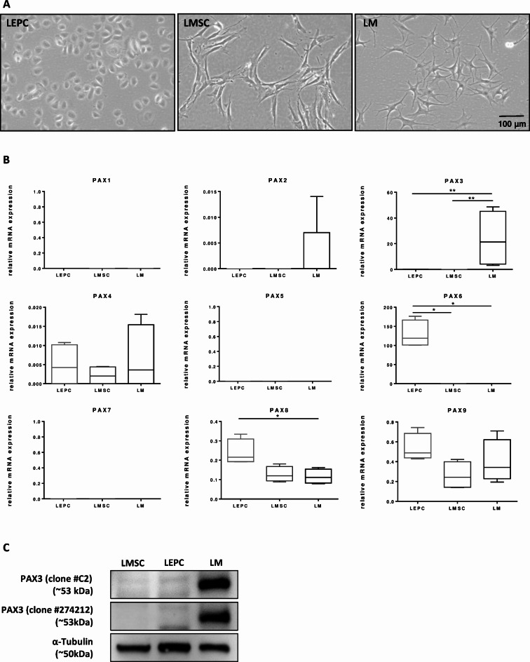
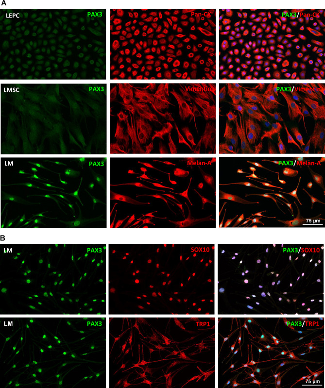
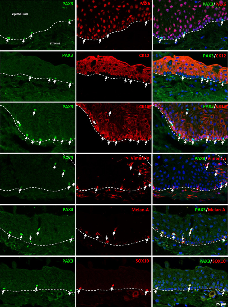
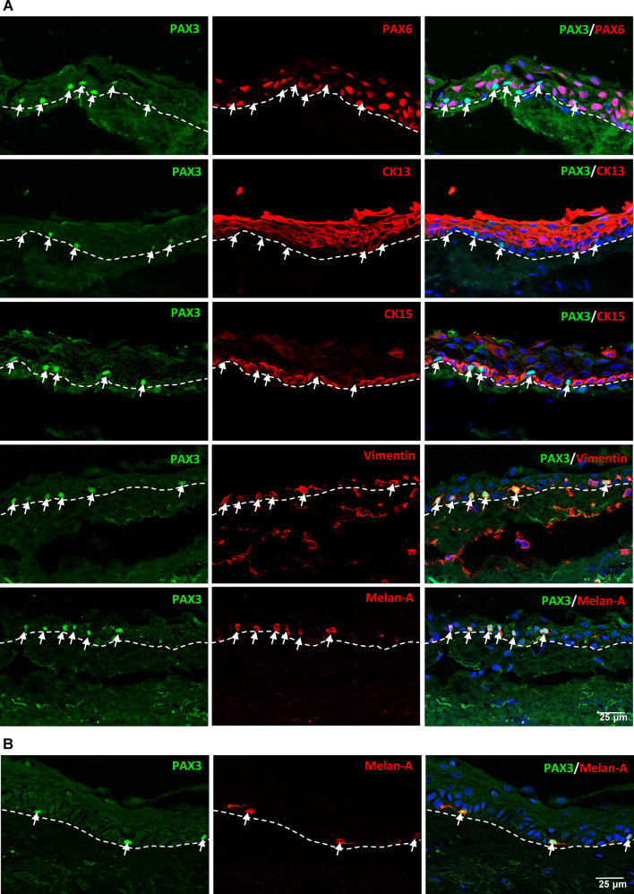
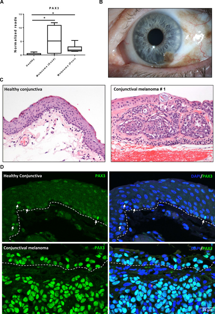
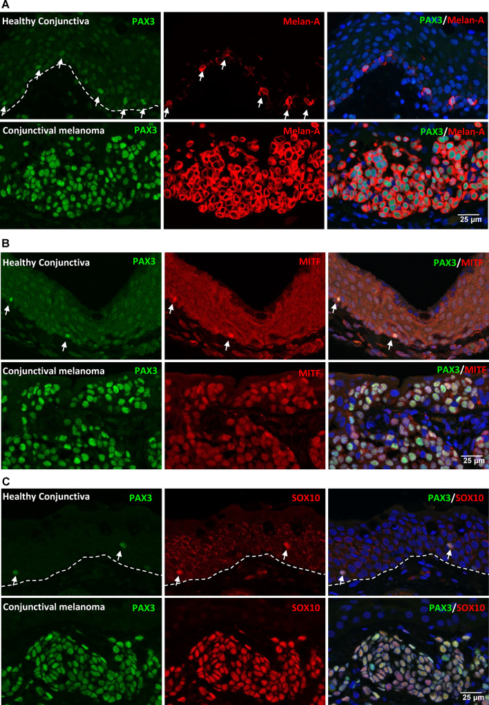
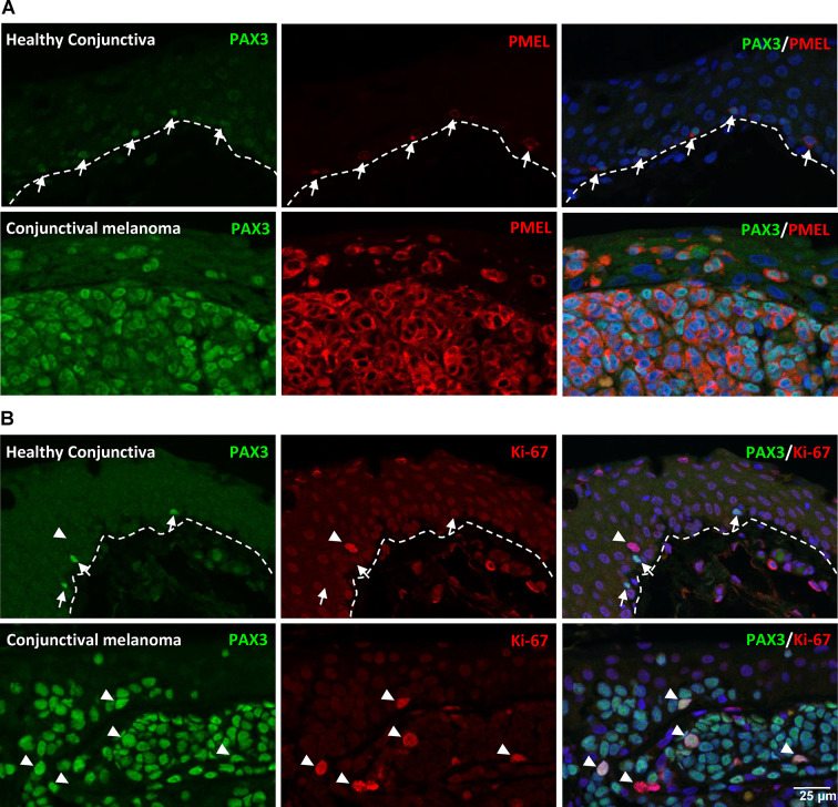
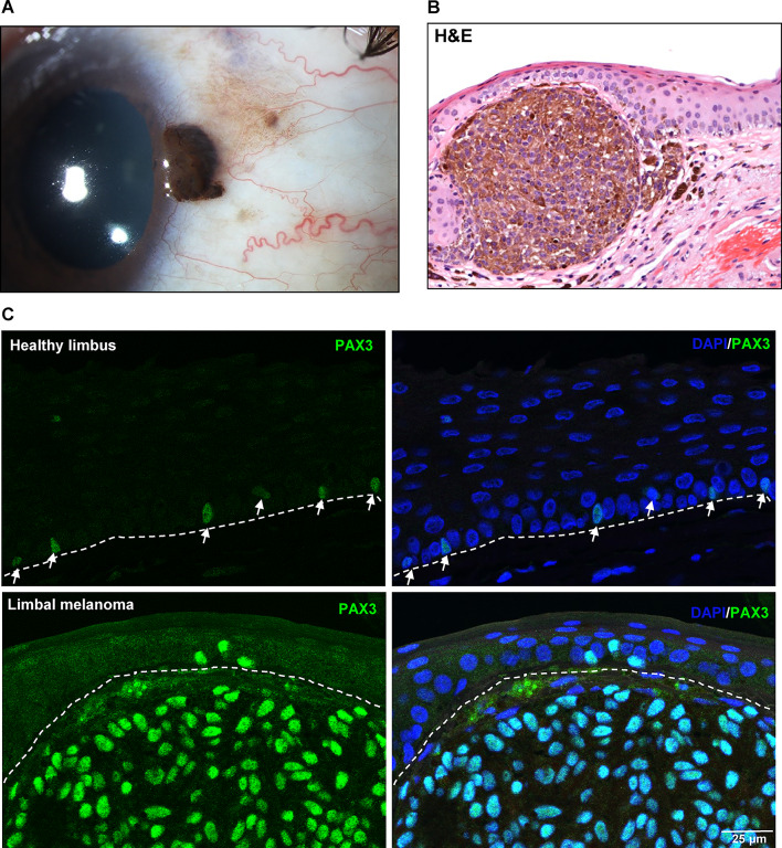
PAX3, a transcription factor essential for neural crest development and melanocyte progenitors, is expressed in various melanocytic tissues. However, its role in ocular surface tissues remains poorly understood. This study investigated the expression patterns of PAX3 in the limbal stem cell niche, specifically in limbal epithelial progenitor cells (LEPC), limbal melanocytes (LM), and limbal mesenchymal stem cells (LMSC). Additionally, PAX3 expression was studied in conjunctival/limbal melanoma specimens. Immunohistochemical analysis revealed predominant PAX3 expression in LM as well in the conjunctival melanocytes, suggesting distinct roles in stem cell regulation and melanocyte maintenance. Notably, PAX3 was significantly upregulated in conjunctival/limbal melanoma tissues compared to healthy counterparts, with expression co-localizing with melanocyte markers (Melan-A, HMB45, SOX10) and the proliferation marker Ki-67 in melanoma cells. These findings suggests that while PAX3 expression is restricted to melanocytes in limbal/conjunctival tissues and its dysregulation may play a crucial role in conjunctival/limbal melanoma development. Further investigation into mechanisms by which PAX3 influences corneal pathophysiology and contributes to conjunctival/limbal melanoma pathogenesis could identify potential therapeutic targets for this aggressive ocular malignancy.

Keywords: Aniridia; Conjunctival melanoma; Limbal epithelial progenitor cells; Limbal melanoma; Limbal niche cells; Limbal stem cell deficiency; Limbal stem cell niche; Limbal stem cells; Melanocytes; Melanoma; Mesenchymal stromal stem cells; PAX3; PAX6.


Conflict of interest statement

Declarations. Competing interests: The authors declare no competing interests. Ethical approval: The Institutional Review Board of the Medical Faculty of the University of Freiburg (25/20) approved experiments. Informed consent: Informed consent to corneal donation had been obtained from the donors or their relatives. Generative AI in scientific writing: During the preparation of this work the authors used Claude AI in order to improve readability and language. After using this tool, the authors reviewed and edited the content as needed and take full responsibility for the content of the publication.

Figures

Fig. 1
Fig. 1
Characterization of limbal cell types and PAX expression. (A) Phase contrast images showing the spindle-shaped morphology, elongated with prominent nucleolus of limbal mesenchymal stromal cells (LMSC); large, flattened, smooth bodies with multiple dendrites of limbal melanocytes (LM); small cuboidal epithelial phenotype of limbal epithelial progenitor cells (LEPC). (B) mRNA expression levels of PAX gene family in cultured LEPC, LMSC and LM. Data are expressed as means (2−ΔCT) ± SEM (n = 5); *p < 0.05; **p < 0.01. Mann-Whitney U test. (C) Western blot analysis of PAX3 expression (using two different clones) in LM, LMSC and LEPC. α-tubulin serves as a loading control. Uncropped versions are available in Suppl. Figure 1C.
Fig. 2
Fig. 2
Immunocytochemical analysis of PAX3 expression in limbal cell types. (A) Immunocytochemical staining of cultured limbal melanocytes (LM) showing the nuclear PAX3 expression co-stained with Melan-A; limbal mesenchymal stromal cells (LMSC) positive for vimentin but negative for PAX3; and limbal epithelial progenitor cells (LEPC) positive for cytokeratins (pan-CK) but negative for PAX3. (B) Double immunostaining of LM revealing co-expression of PAX3 (green) in TRP1+ and SOX10+ cells (red). Nuclear counterstaining with DAPI (blue).
Fig. 3
Fig. 3
PAX3 expression in limbal tissues. Double immunostaining of corneoscleral sections revealing PAX3+ cells (arrows) at the basal layer of limbal epithelium. These cells do not express PAX6, cytokeratin 12 (CK12), or CK15 (red) but do express vimentin (arrow, red) and melanocyte markers Melan-A and SOX10 (red). The dashed line represents the basement membrane. Nuclei are counterstained with DAPI (blue).
Fig. 4
Fig. 4
PAX3 expression in conjunctival and organ-cultured corneoscleral tissues. (A) Double immunostaining of conjunctival tissue sections showing PAX3+ cells (arrows) at the basal layer of conjunctival epithelium. These cells do not express PAX6, cytokeratin 13 (CK13), or CK15 (red) but do express vimentin (arrow, red) and melanocyte markers Melan-A (red) and SOX10. The dashed line represents the basement membrane. (B) Double immunostaining of organ-cultured corneoscleral tissue demonstrating PAX3+ cells (arrows) expressing Melan-A (red). Nuclear counterstaining with DAPI (blue).
Fig. 5
Fig. 5
PAX3 expression in conjunctival melanoma. (A) RNA-sequencing data analysis showing significant upregulation of PAX3 in conjunctival melanoma (both poor and good prognosis) compared to healthy conjunctival tissue. (B) Representative clinical image of conjunctival melanoma: pigmented lesion visible on temporal conjunctiva at 5 o’clock position. (C) Representative histological specimens of normal conjunctiva and melanoma. (D) Immunofluorescent staining of paraffin sections showing strong expression of PAX3 (green) in melanoma compared to healthy tissue. Nuclei are counterstained with DAPI (blue).
Fig. 6
Fig. 6
Melanocyte marker expression in conjunctival melanoma. (A) Double immunofluorescence staining of conjunctival melanoma paraffin sections showing co-expression of PAX3 (green, arrows) and Melan-A (red). Melan-A staining intensity higher in melanoma tissues compared to healthy tissues. (B and C) Expression of melanocyte-specific transcription factors in healthy and melanoma tissues: microphthalmia-associated transcription factor (MITF) is expressed in both no notable difference, while SOX10 is expressed in both with strongest staining observed in melanoma tissues.
Fig. 7
Fig. 7
Pigmentation and proliferation markers in conjunctival melanoma. (A) Double immunofluorescence staining showing expression of the pigmentation marker HMB45: weakly stained or undetectable in healthy conjunctiva, with strong expression in melanoma tissues. (B) Proliferation status of PAX3+ cell: in healthy tissues, PAX3+ cells do not express the proliferation marker Ki-67 (arrows, red), while in melanoma tissues, PAX3+ cells stain positive for Ki-67 (arrowheads), indicating proliferation status.
Fig. 8
Fig. 8
PAX3 expression in limbal melanoma: (A) Representative clinical image of limbal melanoma showing a pigmented lesion. (B) Representative histological specimen of limbal melanoma characterized by the nodular supraepithelial invasion of heavily pigmented melanoma cells. (C) Immunohistochemical analysis revealing the strong expression of PAX3 (green) in limbal melanoma tissues compared to healthy counterpart tissue. Dashed line represents the basement membrane.

Similar articles

Cited by

  • Specific oncogene activation of the cell of origin in mucosal melanoma.
    Babu S, Chen J, Baron CS, Sun K, Robitschek E, McConnell AM, Wu C, Dedeilia A, Sade-Feldman M, Modhurima R, Manos MP, Chen KY, Cox AM, Ludwig CG, Kellis M, Buchbinder EI, Hacohen N, Yang J, Boland GM, Abraham BJ, Liu D, Zon LI, Insco ML. Babu S, et al. Nat Commun. 2025 Jul 22;16(1):6750. doi: 10.1038/s41467-025-61937-1. Nat Commun. 2025. PMID: 40695831 Free PMC article.

References

    1. Gonzalez, G., Sasamoto, Y., Ksander, B. R., Frank, M. H. & Frank, N. Y. Limbal Stem cells: identity, Developmental Origin, and therapeutic potential. Wiley Interdiscip. Rev. Dev. Biol.710.1002/wdev.303 (2018). - PMC - PubMed
    1. Ordonez, P. & Di Girolamo, N. Limbal epithelial stem cells: Role of the Niche microenvironment. Stem Cells. 30, 100–107. 10.1002/stem.794 (2012). - PubMed
    1. Higa, K., Shimmura, S., Miyashita, H., Shimazaki, J. & Tsubota, K. Melanocytes in the corneal Limbus interact with K19-Positive basal epithelial cells. Exp. Eye Res.81, 218–223. 10.1016/j.exer.2005.01.023 (2005). - PubMed
    1. Henkind, P. Migration of limbal melanocytes. Nature214, 1349–1351. 10.1038/2141349b0 (1967). - PubMed
    1. Shimmura, S. & Kawakita, T. Accessory cell populations in the Cornea. Ocul. Surf.4, 74–80. 10.1016/S1542-0124(12)70029-0 (2006). - PubMed

Substances

LinkOut - more resources