

Fabrication and evaluation of 3D printed PLGA/nHA/GO scaffold for bone tissue engineering
- PMID: 40216829
- PMCID: PMC11992093
- DOI: 10.1038/s41598-025-96099-z
Fabrication and evaluation of 3D printed PLGA/nHA/GO scaffold for bone tissue engineering
Abstract
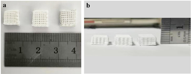
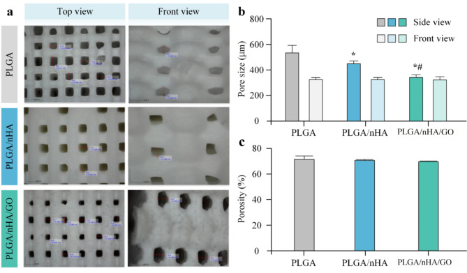
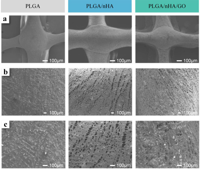
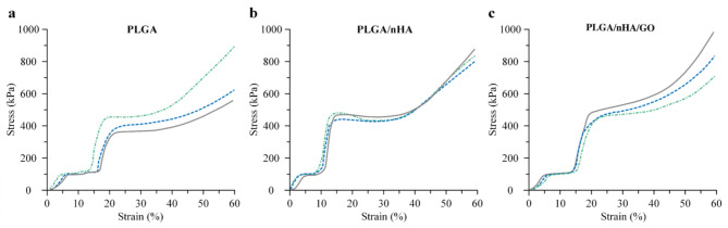
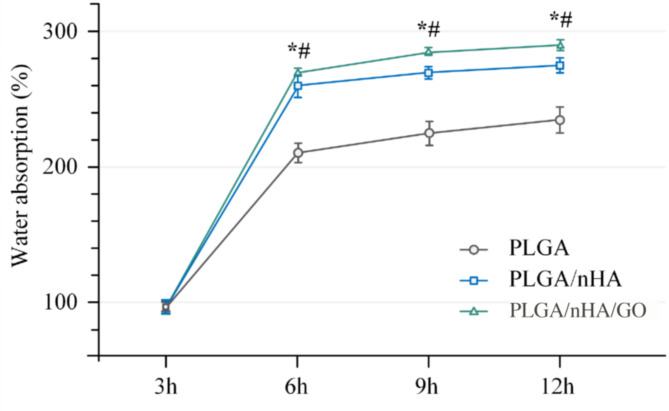
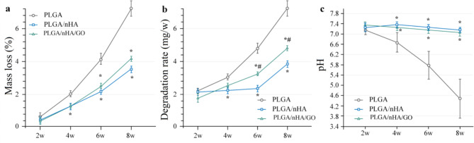
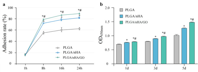
The study aimed to fabricate and evaluate a bone tissue engineering scaffold made from a composite of polylactic-co-glycolic acid (PLGA), nano-hydroxyapatite (nHA), and graphene oxide (GO) using low-temperature 3D printing and freeze-drying techniques. The scaffolds were produced with varying compositions: PLGA alone and in combination with nHA and GO. The macro and microstructure, pore size, porosity, mechanical properties, and in vitro biocompatibility were assessed. Bone marrow mesenchymal stem cells (BMSCs) were co-cultured with the scaffolds to evaluate cell adhesion, proliferation, and cytotoxicity. The PLGA/nHA/GO composite scaffolds exhibited optimal pore size and microtopography, enhanced mechanical properties, excellent water absorption, and appropriate degradability. The co-culture with BMSCs demonstrated improved cell adhesion and proliferation, indicating good biocompatibility. The PLGA/nHA/GO composite scaffolds show potential as a bone tissue engineering material due to their favorable properties and biocompatibility, suggesting their suitability for bone defect repair applications.
Keywords: 3D printing; Bone tissue engineering; Graphene oxide; Nano-hydroxyapatite; Polylactic-co-glycolic acid; Scaffold.
© 2025. The Author(s).
Conflict of interest statement
Declarations. Competing interests: The authors declare no competing interests. Ethics approval and consent to participate: This study was approved by the Biomedical Ethics Committee of Inner Mongolia Medical University (approval number: YKD202405069).
Figures
Similar articles
-
3D-printed nano-hydroxyapatite/poly(lactic-co-glycolic acid) scaffolds with adipose-derived mesenchymal stem cells enhance bone regeneration in rat model of bone defects.J Biomater Appl. 2025 Aug;40(2):284-296. doi: 10.1177/08853282251332050. Epub 2025 Apr 3. J Biomater Appl. 2025. PMID: 40179420
-
Fabrication and in vitro biocompatibility of biomorphic PLGA/nHA composite scaffolds for bone tissue engineering.Mater Sci Eng C Mater Biol Appl. 2014 Mar 1;36:95-101. doi: 10.1016/j.msec.2013.11.047. Epub 2013 Dec 7. Mater Sci Eng C Mater Biol Appl. 2014. PMID: 24433891
-
Effects of Nano-hydroxyapatite/Poly(DL-lactic-co-glycolic acid) Microsphere-Based Composite Scaffolds on Repair of Bone Defects: Evaluating the Role of Nano-hydroxyapatite Content.Artif Organs. 2016 Jul;40(7):E128-35. doi: 10.1111/aor.12741. Artif Organs. 2016. PMID: 27378617
-
Preparation and characterization of PLA/PCL/HA composite scaffolds using indirect 3D printing for bone tissue engineering.Mater Sci Eng C Mater Biol Appl. 2019 Nov;104:109960. doi: 10.1016/j.msec.2019.109960. Epub 2019 Jul 6. Mater Sci Eng C Mater Biol Appl. 2019. PMID: 31500051
-
[CYTOCOMPATIBILITY AND PREPARATION OF BONE TISSUE ENGINEERING SCAFFOLD BY COMBINING LOW TEMPERATURE THREE DIMENSIONAL PRINTING AND VACUUM FREEZE-DRYING TECHNIQUES].Zhongguo Xiu Fu Chong Jian Wai Ke Za Zhi. 2016 Mar;30(3):292-7. Zhongguo Xiu Fu Chong Jian Wai Ke Za Zhi. 2016. PMID: 27281872 Chinese.
Cited by
-
Magnesium phosphate functionalized graphene oxide and PLGA composite matrices with enhanced mechanical and osteogenic properties for bone regeneration.Regen Biomater. 2025 Jul 26;12:rbaf074. doi: 10.1093/rb/rbaf074. eCollection 2025. Regen Biomater. 2025. PMID: 40837704 Free PMC article.
References
-
- Crane, G. M., Ishaug, S. L. & Mikos, A. G. (Nature Publishing Group US New York, (1995).
-
- Dubey, S. K. et al. Uncovering the diversification of tissue engineering on the emergent areas of stem cells, nanotechnology and biomaterials. Curr. Stem Cell Res. Therapy. 15, 187–201 (2020). - PubMed
-
- Middleton, J. C. & Tipton, A. J. Synthetic biodegradable polymers as orthopedic devices. Biomaterials21, 2335–2346 (2000). - PubMed
-
- Wu, X. et al. Preparation of mesoporous nano-hydroxyapatite using a surfactant template method for protein delivery. J. Bionic Eng.9, 224–233 (2012).
-
- Xie, X. H. et al. Structural and degradation characteristics of an innovative porous PLGA/TCP scaffold incorporated with bioactive molecular Icaritin. Biomed. Mater.5, 054109 (2010). - PubMed
MeSH terms
Substances
Grants and funding
- YKD2023BSQD012/Doctoral initiation program of Inner Mongolia Medical University
- YKD2024MS006/Research Project of InnerMongolia Medical University
- 2024SZY0127/The central government guides local funds for scientific and technological development
- NMGIRT2419/Innovation Team Development Plan for Higher Education Institutions in Inner Mongolia Autonomous Region
- 2020/"Grassland Talent" project for youth innovation and entrepreneurship talent in Inner Mongolia Autonomous Region (2020),
- 2021/Science research project of Inner Mongolia Autonomous Region Mongolian Medicine and Pharmaceutical Collaborative Innovation Center in 2021
- YKD2021ZD001/Inner Mongolia Medical University 2021 annual school-level research key project
- NMGIRT2227/Inner Mongolia Higher Education Innovation Team Development Program
- 2023YFHH0003/Inner Mongolia Autonomous Region key research & development & achievement transformation plan ( 2023 Science and Technology to support the Yellow River Basin ecological protection and high-quality development) project
- NJYT24031/Yong Scientific and Technological Talent Program of Institutes of Higher Education of Inner Mongolia Education Department
LinkOut - more resources
Full Text Sources
