

Predicting PD-L1 status in NSCLC patients using deep learning radiomics based on CT images
- PMID: 40216830
- PMCID: PMC11992188
- DOI: 10.1038/s41598-025-91575-y
Predicting PD-L1 status in NSCLC patients using deep learning radiomics based on CT images
Abstract
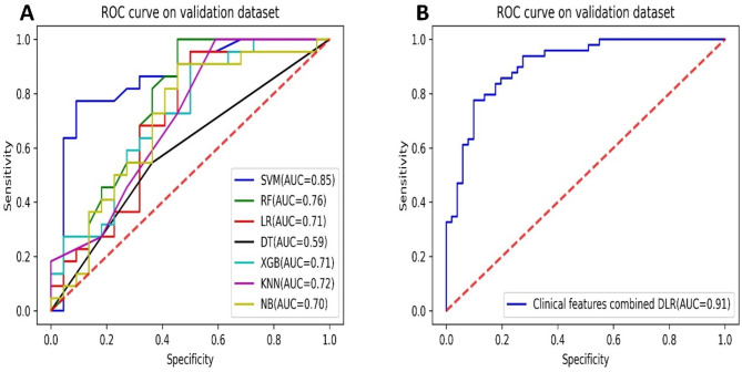
Radiomics refers to the utilization of automated or semi-automated techniques to extract and analyze numerous quantitative features from medical images, such as computerized tomography (CT) or magnetic resonance imaging (MRI) scans. This study aims to develop a deep learning radiomics (DLR)-based approach for predicting programmed death-ligand 1 (PD-L1) expression in patients with non-small cell lung cancer (NSCLC). Data from 352 NSCLC patients with known PD-L1 expression were collected, of which 48.29% (170/352) were tested positive for PD-L1 expression. Tumor regions of interest (ROI) were semi-automatically segmented based on CT images, and DL features were extracted using Residual Network 50. The least absolute shrinkage and selection operator (LASSO) algorithm was used for feature selection and dimensionality reduction. Seven algorithms were used to build models, and the most optimal ones were identified. A combined model integrating DLR with clinical data was also developed. The predictive performance of each model was evaluated using the area under the curve (AUC) of the receiver operating characteristic (ROC) curve analysis. The DLR model, based on CT images, demonstrated an AUC of 0.85 (95% confidence interval (CI), 0.82-0.88), sensitivity of 0.80 (0.74-0.85), and specificity of 0.73 (0.70-0.77) for predicting PD-L1 status. The integrated model exhibited superior performance, with an AUC of 0.91 (0.87-0.95), sensitivity of 0.85 (0.82-0.89), and specificity of 0.75 (0.72-0.80). Our findings indicate that the DLR model holds promise as a valuable tool for predicting the PD-L1 status in patients with NSCLC, which can greatly assist in clinical decision-making and the selection of personalized treatment strategies.
Keywords: Deep learning; Non-small cell lung cancer (NSCLC); Programmed death-ligand 1(PD-L1)..
© 2025. The Author(s).
Conflict of interest statement
Declarations. Ethics approval: The studies involving human participants underwent a thorough review and received approval from the Institutional Review Committee of the First Affiliated Hospital of Shandong First Medical University (Jinan, China). Consent: The informed consent was waived by the First Affiliated Hospital of Shandong First Medical University. Competing interests: The authors declare no competing interests.
Figures


Similar articles
-
CT-based deep learning radiomics biomarker for programmed cell death ligand 1 expression in non-small cell lung cancer.BMC Med Imaging. 2024 Jul 31;24(1):196. doi: 10.1186/s12880-024-01380-8. BMC Med Imaging. 2024. PMID: 39085788 Free PMC article.
-
Radiomics study for predicting the expression of PD-L1 in non-small cell lung cancer based on CT images and clinicopathologic features.J Xray Sci Technol. 2020;28(3):449-459. doi: 10.3233/XST-200642. J Xray Sci Technol. 2020. PMID: 32176676
-
Radiomics Models Derived From Arterial-Phase-Enhanced CT Reliably Predict Both PD-L1 Expression and Immunotherapy Prognosis in Non-small Cell Lung Cancer: A Retrospective, Multicenter Cohort Study.Acad Radiol. 2025 Jan;32(1):493-505. doi: 10.1016/j.acra.2024.07.028. Epub 2024 Jul 31. Acad Radiol. 2025. PMID: 39084935
-
Predictive value of machine learning for PD-L1 expression in NSCLC: a systematic review and meta-analysis.World J Surg Oncol. 2025 May 22;23(1):199. doi: 10.1186/s12957-025-03847-6. World J Surg Oncol. 2025. PMID: 40405177 Free PMC article.
-
Predictive value of 18F-fluorodeoxyglucose positron emission tomography/computed tomography for PD-L1 expression in non-small cell lung cancer: A systematic review and meta-analysis.Thorac Cancer. 2020 Nov;11(11):3260-3268. doi: 10.1111/1759-7714.13664. Epub 2020 Sep 20. Thorac Cancer. 2020. PMID: 32951338 Free PMC article.
Cited by
-
Explanation and Elaboration with Examples for METRICS (METRICS-E3): an initiative from the EuSoMII Radiomics Auditing Group.Insights Imaging. 2025 Aug 13;16(1):175. doi: 10.1186/s13244-025-02061-y. Insights Imaging. 2025. PMID: 40802002 Free PMC article.
References
-
- Loh, J. et al. Management of oncogene driven locally advanced unresectable non-small cell lung cancer. Expert Rev. Anticancer Ther.23(9), 913–926 (2023). - PubMed
-
- Ettinger, D. S. et al. NCCN guidelines insights: non-small cell lung cancer, version 2.2021. J. Natl. Compr. Canc Netw.19(3), 254–266 (2021). - PubMed
MeSH terms
Substances
Grants and funding
LinkOut - more resources
Full Text Sources
Medical
Research Materials
