. 2025 Apr 11;15(1):12492.
doi: 10.1038/s41598-025-97831-5.

Mechanism of dracorhodin in accelerating diabetic foot ulcer healing via the Nrf2 pathway, a network pharmacology, molecular docking and experimental validation

Affiliations

Mechanism of dracorhodin in accelerating diabetic foot ulcer healing via the Nrf2 pathway, a network pharmacology, molecular docking and experimental validation

Guangjun Tang et al. Sci Rep. .

Abstract

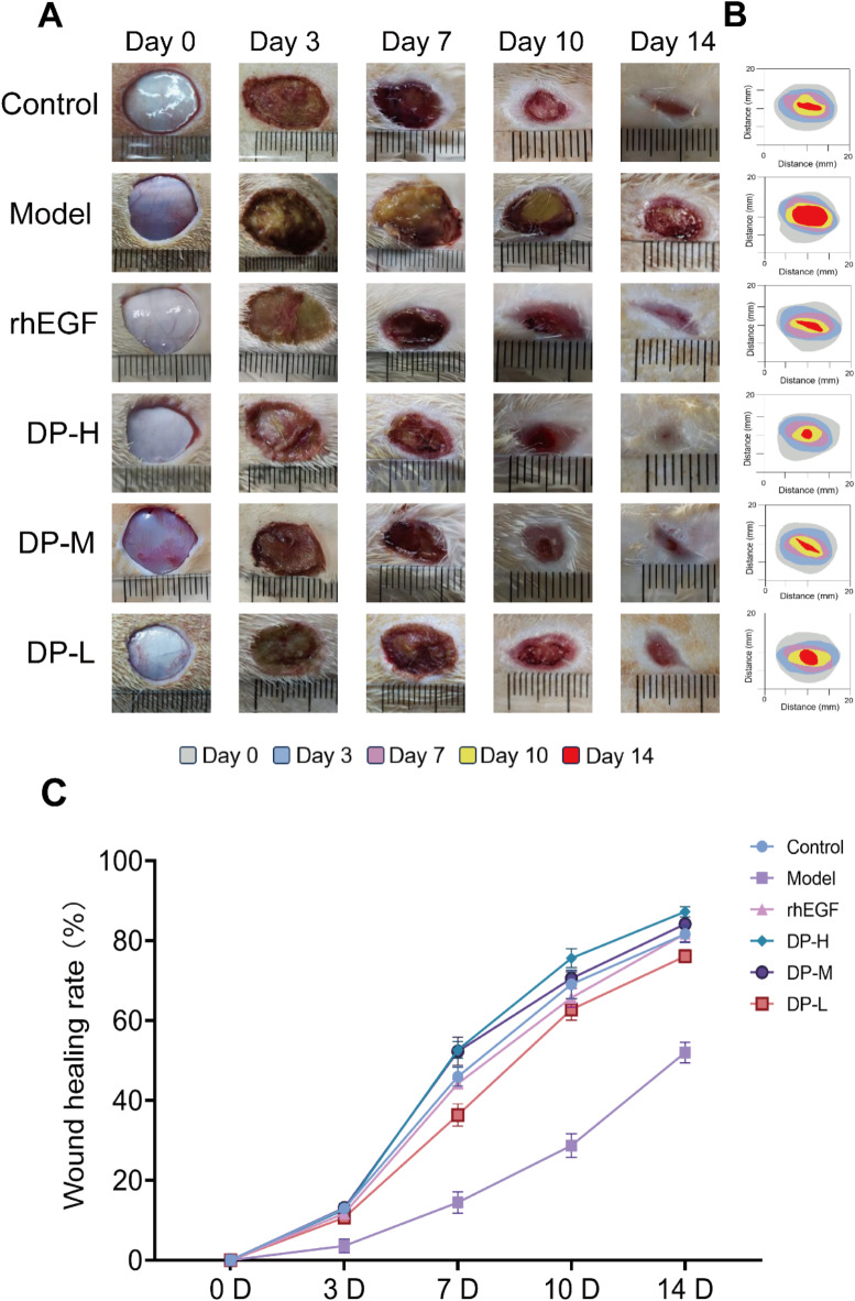
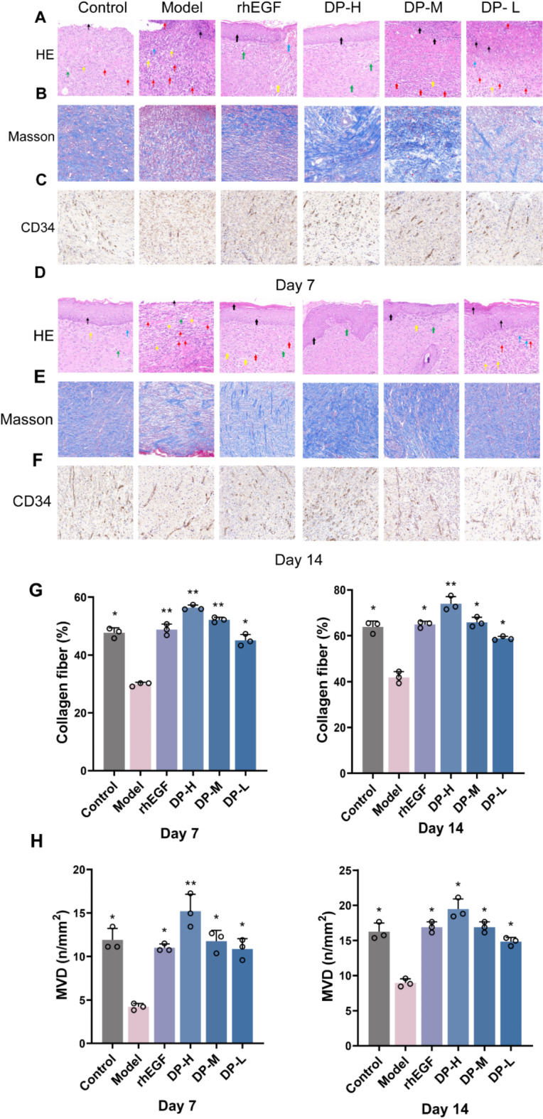
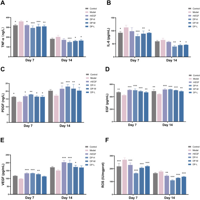
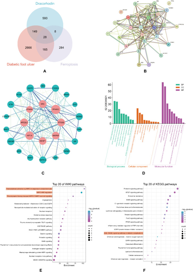
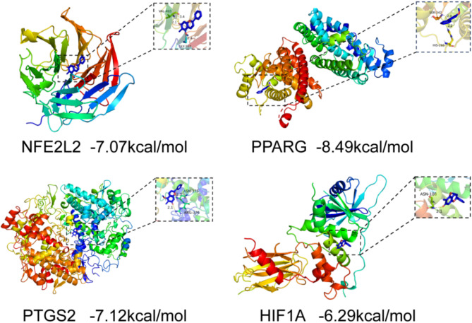
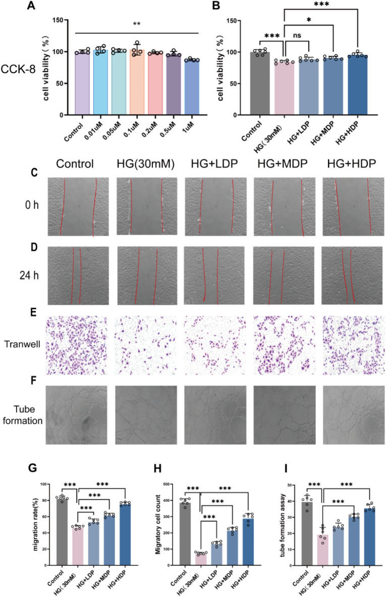
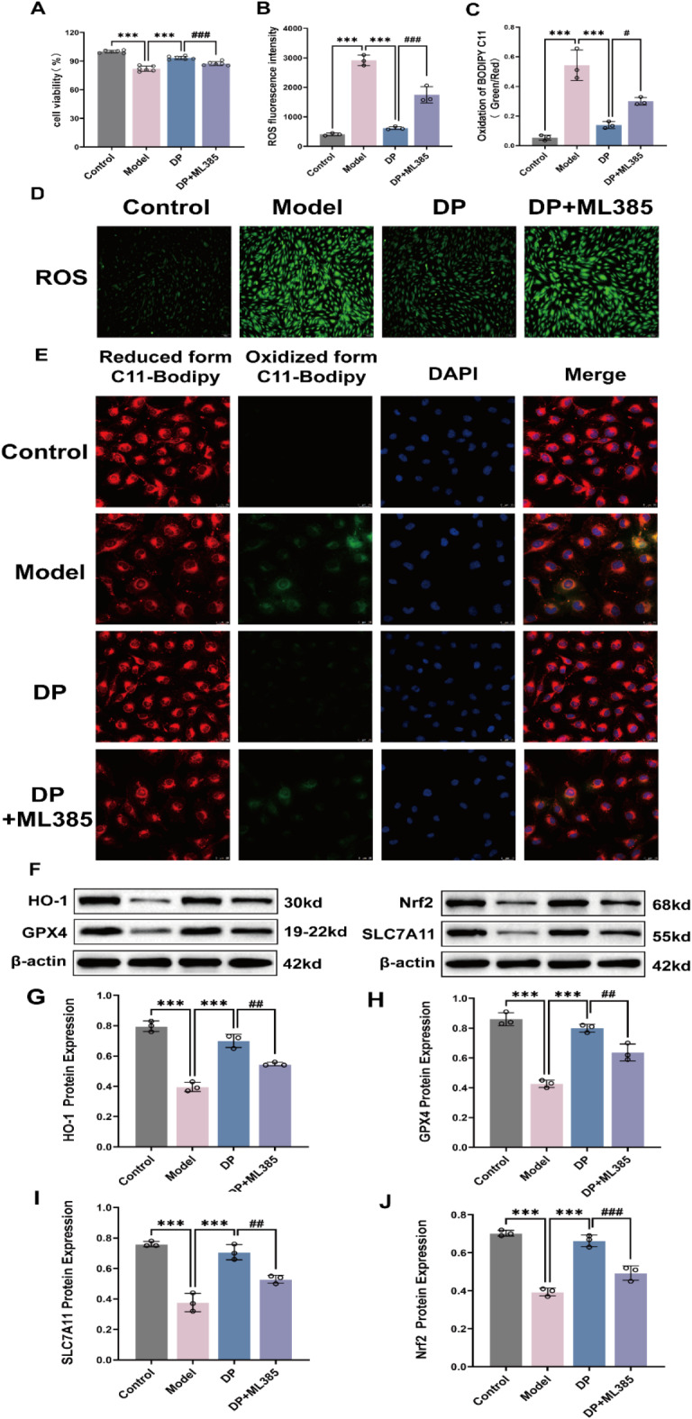
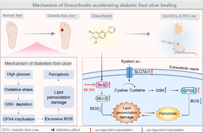
Delayed wound healing in diabetic foot ulcer (DFU) is a major cause of amputations, with ferroptosis impeding recovery. Dracorhodin (DP), a flavonoid from Dragon's Blood, has shown anti-inflammatory and wound-healing properties, though its molecular mechanisms is unclear. This study investigates DP's role in DFU treatment through bioinformatics and experimental approaches. A rat model of DFU was created with a high-fat/high-glucose diet and streptozotocin (STZ) induction, and wound healing was monitored after applying varying DP doses. Histopathological analysis and ELISA assessed tissue changes, inflammatory markers, and growth factors. Network pharmacology and molecular docking were used to identify core targets and pathways, while human umbilical vein endothelial cells (HUVECs) were used for in vitro testing. The results demonstrated that DP accelerated wound healing in DFU rats in a dose-dependent manner by enhancing collagen synthesis, angiogenesis, and growth factor levels, while simultaneously reducing inflammation and ROS levels. Network pharmacology and molecular docking analyses identified the Nrf2-mediated ferroptosis pathway as a potential key mechanism underlying DP's therapeutic effects in DFU. In vitro experiments further revealed that DP improved cell viability and migration, while decreasing ROS and lipid peroxidation levels, effects attributed to Nrf2 pathway activation. These outcomes were significantly attenuated by the Nrf2 inhibitor ML385. In conclusion, DP promotes DFU healing via activation of the Nrf2 pathway and inhibition of ferroptosis.

Keywords: Diabetic foot ulcer; Dracorhodin; Ferroptosis; Nrf2 pathway.


Conflict of interest statement

Declarations. Competing interests: The authors declare no competing interests.

Figures

Fig. 1
Fig. 1
DP accelerates wound healing in DFU rats. (A) Representative images of wound area healing at various time points in DFU rats. (B) DFU rat wound healing simulation diagram. (C) Wound healing rates (%) at day 0, 3, 7, 10, and 14 across different groups.
Fig. 2
Fig. 2
Topical application of DP significantly promotes collagen fiber deposition and neovascularization in DFU rat models, while reducing inflammatory cell infiltration and increasing MVD. (A,D) HE staining of DFU rat wounds on day 7 and 14, scale bar = 50 μm. Black arrows denote the loss of epidermal layer integrity within the field of view, yellow arrows indicate collagen fiber content in the dermis, green arrows highlight newly formed blood vessels, red arrows represent inflammatory cell infiltration, and blue arrows mark areas of bleeding. (B,E) Masson staining of DFU rat wounds on day 7 and 14, scale bar = 50 μm. (C,F) CD34 immunohistochemistry of DFU rat wounds on day 7 and 14, scale bar = 50 μm. (G) Quantitative analysis of collagen fiber occupancy by Masson staining of DFU rat wounds on day 7 and 14. (H) Quantitative analysis of MVD in DFU rat wounds on days 7 and 14. Data were expressed as mean ± SD (n = 3). *P<0.05, **P<0.01, ***P<0.001 compared with the model group.
Fig. 3
Fig. 3
Results of serum Elisa on day 7 and 14 in DFU rats. (A) TNF-α results. (B) IL-6 results. (C) PDGF results. (D) EGF results. (E) VEGF results. (F) ROS results. Data were expressed as mean ± SD (n = 3). *P<0.05, **P<0.01, ***P<0.001 compared with the model group.
Fig. 4
Fig. 4
Pharmacological analysis of the DP-DFU-Ferroptosis network. (A) Venny figure. (B) PPI network analysis. (C) Key components of the DP-DFU-Ferroptosis core target network. (D) GO enrichment analysis. (E) WIKI enrichment analysis. (F) KEGG enrichment analysis.
Fig. 5
Fig. 5
Visualization of docking between DP and core target molecules (binding energy ≤ − 5.0 kcal/mol).
Fig. 6
Fig. 6
Effects of DP on HUVECs with high glucose damage. (A) Screening of DP working concentrations by CCK-8 method. (B) CCK-8 assay for cell viability. (C,D,G) Scratch assay and statistical analysis. (E,H) Transwell assay and statistical analysis. (F,I) Tube formation assay and statistical analysis. Data were expressed as mean ± SD (n = 3). *P<0.05, **P<0.01, ***P<0.001 compared with the model group, #P<0.05, ##P<0.01, ###P<0.001 compared with the DP group.
Fig. 7
Fig. 7
DP activates the Nrf2 pathway to inhibit ferroptosis to repair high glucose-damaged HUVECs. (A) Cell viability assay of high glucose-damaged HUVECs after treatment with DP and ML385. (B,D) Immunofluorescence and statistical analysis of ROS (scale bar = 100 μm). (C,E) Lipid peroxidation assay (scale bar = 25 μm). (FJ) WB detection of Nrf2 pathway-related protein expression and quantitative analysis. Data were expressed as mean ± SD (n = 3). *P<0.05, **P<0.01, ***P<0.001, #P<0.05, ##P<0.01, ###P<0.001 compared with the DP group. Original blots are presented in Supplementary Fig. 1–6.
Fig. 8
Fig. 8
DP accelerates diabetic foot ulcer healing by alleviating ferroptosis via the Nrf2 pathway activation.

Similar articles

Cited by

References

    1. GBD 2021 Diabetes Collaborators. Global, regional, and National burden of diabetes from 1990 to 2021, with projections of prevalence to 2050: a systematic analysis for the global burden of disease study 2021. Lancet402(10397), 203–234 (2023). - PMC - PubMed
    1. International Diabetes Federation. The Diabetic Foot. Brussels, Belgium & Federation, I. D. https://www.idf.org/our-activities/care-prevention/diabetic-foot.html (Accessed 1 Aug 2022) (2020).
    1. Yadav, J. P. et al. Phytoconstituents as modulators of NF-κB signalling: investigating therapeutic potential for diabetic wound healing. Biomed. Pharmacother. 177, 117058 (2024). - PubMed
    1. Deng, P. et al. Worldwide research trends on diabetic foot ulcer (2004–2020): Suggestions for researchers. J. Diabetes Res.2022, 7991031. (2022). - PMC - PubMed
    1. Chen, L., Sun, S., Gao, Y. & Ran, X. Global mortality of diabetic foot ulcer: A systematic review and meta-analysis of observational studies. Diabetes Obes. Metab.25(1), 36–45 (2023). - PubMed

LinkOut - more resources