Demethylating drugs alter protoplast development, regeneration, and the genome stability of protoplast-derived regenerants of cabbage
- PMID: 40217153
- PMCID: PMC11987290
- DOI: 10.1186/s12870-025-06473-2
Demethylating drugs alter protoplast development, regeneration, and the genome stability of protoplast-derived regenerants of cabbage
Abstract
Background: Methylation is a major DNA modification contributing to the epigenetic regulation of nuclear gene expression and genome stability. DNA methyltransferases (DNMT) inhibitors are widely used in epigenetic and cancer research, but their biological effects and the mechanisms of their action are not well recognized in plants. This research focuses on comparing the effects of two DNMT inhibitors, namely 5-azacytidine (AZA) and zebularine (ZEB), on cellular processes, including organogenesis in vitro. Protoplasts are a unique single-cell system to analyze biological processes in plants; therefore in our study, both inhibitors were applied to protoplast culture medium or the medium used for the regeneration of protoplast-derived calluses.
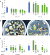
Results: AZA induced a dose-dependent reduction in protoplast viability, delayed cell wall reconstruction, and reduced mitotic activity, while ZEB in low concentration (2.5 µM) promoted mitoses and stimulated protoplast-derived callus development. The higher effectiveness of shoot regeneration was observed when drugs were applied directly to protoplasts compared to protoplast-derived callus treatments. Our findings reveal that both drugs affected the genome stability of the obtained regenerants by inducing polyploidization. Both drugs induced hypomethylation and modulated the distribution patterns of methylated DNA in the protoplast-derived callus.
Conclusion: AZA was more toxic to plant protoplasts compared to ZEB. Both inhibitors affect the ploidy status of protoplast-derived regenerants. A comparison of the data on global methylation levels with the regeneration efficiency suggests that organogenesis in cabbage is partially controlled by variations in DNA methylation levels.
Keywords: Cabbage; DNA hypomethylation; Methyltransferase inhibitors; Mitotic divisions; Ploidy; Protoplasts; Regeneration.
© 2025. The Author(s).
Conflict of interest statement
Declarations. Ethics approval and consent to participate: Not applicable. Consent for publication: Not applicable. Competing interests: The authors declare no competing interests. Clinical trial number: Not applicable.
Figures


Similar articles
-
Development of an optimized protocol for protoplast-to-plant regeneration of selected varieties of Brassica oleracea L.BMC Plant Biol. 2024 Dec 30;24(1):1279. doi: 10.1186/s12870-024-06005-4. BMC Plant Biol. 2024. PMID: 39736572 Free PMC article.
-
Sugarbeet (Beta vulgaris L.): shoot regeneration from callus and callus protoplasts.Planta. 2003 Jul;217(3):374-81. doi: 10.1007/s00425-003-1006-7. Epub 2003 Mar 14. Planta. 2003. PMID: 14520564
-
Effects of TSA, NaB, Aza in Lactuca sativa L. protoplasts and effect of TSA in Nicotiana benthamiana protoplasts on cell division and callus formation.PLoS One. 2023 Feb 24;18(2):e0279627. doi: 10.1371/journal.pone.0279627. eCollection 2023. PLoS One. 2023. PMID: 36827385 Free PMC article.
-
Efficient regeneration of protoplasts from Solanum lycopersicum cultivar Micro-Tom.Biol Methods Protoc. 2024 Feb 6;9(1):bpae008. doi: 10.1093/biomethods/bpae008. eCollection 2024. Biol Methods Protoc. 2024. PMID: 38414647 Free PMC article. Review.
-
DNA methyltransferases as targets for cancer therapy.Drugs Today (Barc). 2007 Jun;43(6):395-422. doi: 10.1358/dot.2007.43.6.1062666. Drugs Today (Barc). 2007. PMID: 17612710 Review.
References
-
- Leister RT, Katagiri F. A resistance gene product of the nucleotide binding site-leucine rich repeats class can form a complex with bacterial avirulence proteins in vivo. Plant J. 2000;22:345–54. - PubMed
-
- Reyna-Llorens I, Ferro-Costa M, Burgess SJ. Plant protoplasts in the age of synthetic biology. J Exp Bot. 2023. 10.1093/jxb/erad172. - PubMed
-
- Vu HM, Lee JY, Kim Y, Park S, Izaguirre F, Lee J, Lee J-H, Jo M, Woo RH, Kim JY, Lim PO, Kim MS. Exploring the feasibility of a single-protoplast proteomic analysis. J Anal Sci Technol. 2024. 10.1186/s40543-024-00457-x.
MeSH terms
Substances
Grants and funding
LinkOut - more resources
Full Text Sources

