

Histological and immunohistochemical analysis of human periapical lesions: a study of TGF-β1 and CD68 markers
- PMID: 40217194
- PMCID: PMC11987189
- DOI: 10.1186/s12903-025-05845-2
Histological and immunohistochemical analysis of human periapical lesions: a study of TGF-β1 and CD68 markers
Abstract
Background: Various inflammatory and anti-inflammatory mediators, along with diverse cell types, are implicated in the development and progression of periapical lesions. This work aimed to assess the immuno-expression of transforming growth factor-beta 1 (TGF-β1) and CD68 (a macrophage marker), elucidating their roles and potential correlations. Additionally, histological analysis was conducted to evaluate the intensity of inflammatory infiltrates in chronic periapical lesion samples.
Methods: Tissue samples from fifty individuals with chronic periapical lesions [25 radicular cysts (RCs) and 25 periapical granulomas (PGs)] were obtained, along with control samples from four healthy third molars' dental pulp. Histological examination and inflammatory infiltrate categorization were performed. Immunohistochemical analysis of TGF-β1 and CD68 markers, along with morphometric assessment, were conducted.
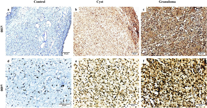
Results: The control group displayed normal, inflammation-free pulp tissues, while intense inflammation was observed in PGs and RCs (Score 4 and 3, respectively) dominated by macrophages, plasma cells, and lymphocytes. Immunohistochemistry showed higher TGF-β1 and CD68 expression in PGs and RCs versus control (P < 0.001). Moreover, PGs exhibited greater TGF-β1 and CD68 expression than RCs (P < 0.001). However, a negative relationship was detected between the 2 markers (P < 0.05).
Conclusions: This study highlighted varying expressions of TGF-β1 and CD68 in PGs and RCs, indicating their potential roles in lesion pathology. However, a negative correlation between these markers was observed. Accordingly, their precise role in periapical lesion progression and repair requires further investigation.
Keywords: Immunohistochemistry; Inflammation; Macrophages; Periapical granuloma; Radicular cyst.
© 2025. The Author(s).
Conflict of interest statement
Declarations. Consent for publication: Not applicable. Competing interests: The authors declare no competing interests. Ethical approval and consent to participate: Ethical approval for this study was granted by the Institutional Ethical Committee of the Faculty of Dentistry, Cairo University (Approval no. 16/10/21), in line with the guidelines set forth in the Declaration of Helsinki. Informed consent was acquired from all participating patients before sample collection, following a thorough explanation of the study’s goals, procedures, and any potential discomforts and risks.
Figures


Similar articles
-
Immunoexpression of interleukin 17, transforming growth factor β1, and forkhead box P3 in periapical granulomas, radicular cysts, and residual radicular cysts.J Endod. 2013 Aug;39(8):990-4. doi: 10.1016/j.joen.2013.04.028. Epub 2013 May 21. J Endod. 2013. PMID: 23880265
-
Macrophage polarization in human periapical lesions in relation to histopathological diagnosis, clinical features and lesion volume: An ex vivo study.Int Endod J. 2024 Dec;57(12):1829-1847. doi: 10.1111/iej.14138. Epub 2024 Sep 2. Int Endod J. 2024. PMID: 39222032
-
Immunohistochemical expression of TGF-β1 and MMP-9 in periapical lesions.Braz Oral Res. 2017 Jul 3;31:e51. doi: 10.1590/1807-3107BOR-2017.vol31.0051. Braz Oral Res. 2017. PMID: 28678970
-
Macrophages subpopulations in chronic periapical lesions according to clinical and morphological aspects.Braz Oral Res. 2019 May 27;33:e047. doi: 10.1590/1807-3107bor-2019.vol33.0047. Braz Oral Res. 2019. PMID: 31141038
-
Accuracy of Ultrasound Imaging in the Differential Diagnosis of Inflammatory Radicular Cyst and Periapical Granuloma: A Systematic Review and Meta-Analysis of Operative Characteristics.Eur Endod J. 2025 Mar;10(2):104-115. doi: 10.14744/eej.2024.84755. Eur Endod J. 2025. PMID: 40143569 Free PMC article.
References
-
- Braz-Silva PH, Bergamini ML, Mardegan AP, De Rosa CS, Hasseus B, Jonasson P. Inflammatory profile of chronic apical periodontitis: a literature review. Acta Odontol Scand. 2019;77(3):173–80. 10.1080/00016357.2018.1521005. - PubMed
-
- Ricucci D, Bergenholtz G. Histologic features of apical periodontitis in human biopsies. Endod Top. 2005;8(1):68–87. 10.1111/j.1601-1546.2004.00097.x.
-
- Koivisto T, Bowles WR, Rohrer M. Frequency and distribution of radiolucent jaw lesions: a retrospective analysis of 9,723 cases. J Endod. 2012;38:729–32. 10.1016/j.joen.2012.02.028. - PubMed
MeSH terms
Substances
LinkOut - more resources
Full Text Sources
