

Operative treatment of severe scoliosis and pelvic obliquity in patients with spinal muscular atrophy: assessment of outcomes and complications
- PMID: 40217273
- PMCID: PMC11992721
- DOI: 10.1186/s13023-025-03682-8
Operative treatment of severe scoliosis and pelvic obliquity in patients with spinal muscular atrophy: assessment of outcomes and complications
Abstract
Background: Few reports exist that focus on patients with spinal muscular atrophy (SMA) and severe spinal deformity. In this study, we aimed to report surgical outcomes and complications for SMA patients with severe scoliosis and pelvic obliquity.
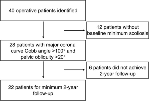
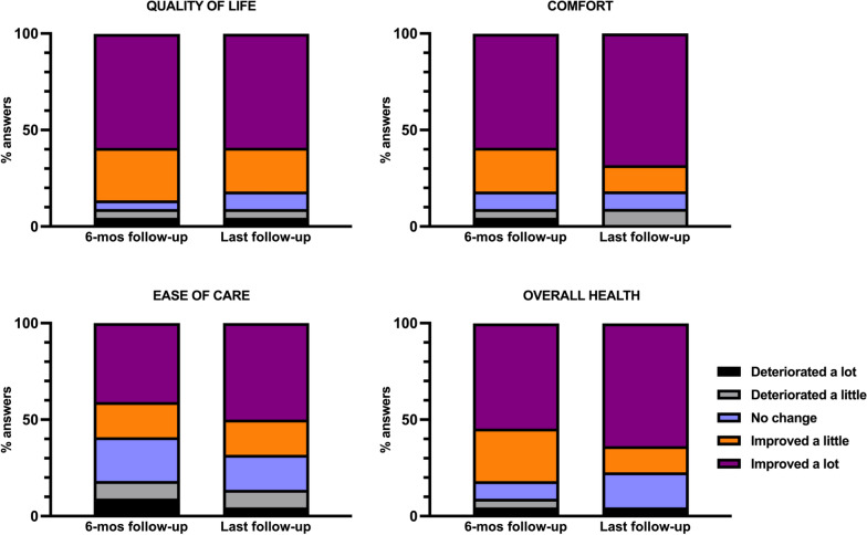
Methods: A retrospective review of data on operatively treated SMA patients with severe scoliosis and pelvic obliquity (minimum major coronal curve Cobb angle > 100° and pelvic obliquity > 20°) was performed. Radiography findings, pulmonary function, motor status, the sitting function score, and perioperative and postoperative complications were the main clinical outcomes examined. Muscular dystrophy spine questionnaire (MDSQ) responses and caregiver responses to four anchor questions (quality of life/comfort/ease of care/overall health) using Likert scales were recorded.
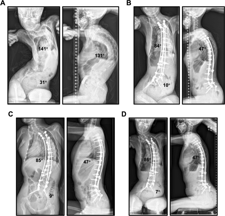
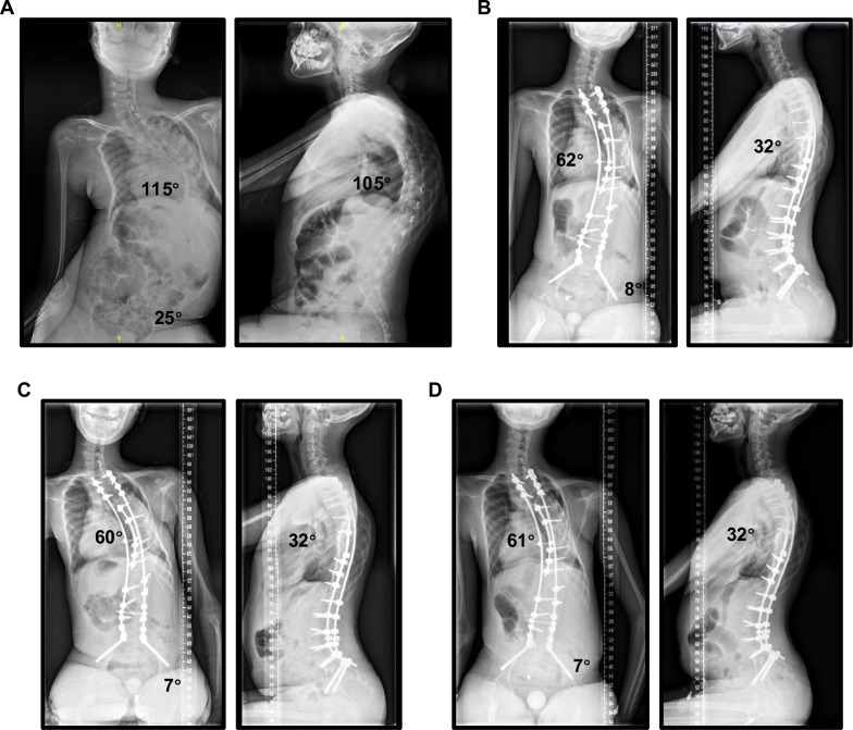
Results: Of 28 consecutive patients, 22 (79%) completed the minimum 2-year follow-up (mean age at surgery = 16.1, 68% female). The mean follow-up duration was 40.3-mo. All patients underwent one-stage posterior spinal fusion (PSF) with pelvic fixation. Radiographic measurements (main coronal curve, kyphosis, pelvic obliquity) were significantly corrected (all p < 0.001) and were maintained at the last follow-up. The mean forced vital capacity (FVC) remained stable during follow-up, with 50% of patients showing improvement. The percentage of patients who could sit independently increased significantly from 22.7% preoperatively to 77.3% postoperatively (p < 0.001). The total sitting-related MDSQ score significantly increased from 8.5 to 12.5 at 6 months postoperatively, and to 15.0 at the last follow-up (p < 0.001). Six instances of complications (two instances each of pneumonia, epiglottic edema, and delayed wound healing) occurred perioperatively in six patients (27.3%), but no surgical intervention was required.
Conclusion: Operative treatment significantly improved radiographic parameters and sitting function and maintained pulmonary function without serious complications in SMA patients with severe scoliosis and pelvic obliquity.
Keywords: Clinical outcome; Pelvic fixation; Posterior spinal fixation; Scoliosis; Spinal muscular atrophy.
© 2025. The Author(s).
Conflict of interest statement
Declarations. Ethics approval: The Institutional Board of Peking union medical college hospital approved this study (IRB: S-K1863), and all patients provided consent for study participation. Consent for publication: Not applicable. Competing interests: The authors declare that they have no competing interests.
Figures
Similar articles
-
Surgical correction of spinal deformity in patients with congenital muscular dystrophy.J Orthop Sci. 2010 Jul;15(4):493-501. doi: 10.1007/s00776-010-1486-9. Epub 2010 Aug 19. J Orthop Sci. 2010. PMID: 20721717
-
Minimum 5-year radiographic results of long scoliosis fusion in juvenile spinal muscular atrophy patients: major curve progression after instrumented fusion.J Pediatr Orthop. 2011 Jul-Aug;31(5):480-8. doi: 10.1097/BPO.0b013e318220ba33. J Pediatr Orthop. 2011. PMID: 21654453
-
Segmental Pedicle Screw Instrumentation and Fusion Only to L5 in the Surgical Treatment of Flaccid Neuromuscular Scoliosis.Spine (Phila Pa 1976). 2018 Mar 1;43(5):331-338. doi: 10.1097/BRS.0000000000000996. Spine (Phila Pa 1976). 2018. PMID: 29095413
-
Survival of patients with Duchenne muscular dystrophy who underwent spinal deformity correction.Dev Med Child Neurol. 2024 Feb;66(2):187-194. doi: 10.1111/dmcn.15711. Epub 2023 Jul 21. Dev Med Child Neurol. 2024. PMID: 37482906 Review.
-
[Characteristics in the treatment of scoliosis in muscular diseases].Z Orthop Ihre Grenzgeb. 1997 Mar-Apr;135(2):95-105. doi: 10.1055/s-2008-1039563. Z Orthop Ihre Grenzgeb. 1997. PMID: 9214180 Review. German.
References
-
- Wijngaarde CA, et al. Natural course of scoliosis and lifetime risk of scoliosis surgery in spinal muscular atrophy. Neurology. 2019;93(2):e149–58. - PubMed
-
- Coratti G, et al. Early treatment of type II SMA slows rate of progression of scoliosis. J Neurol Neurosurg Psychiatr. 2024;95(3):235–40. - PubMed
-
- Chandran S, et al. Early treatment of scoliosis with growing rods in children with severe spinal muscular atrophy: a preliminary report. J Pediatr Orthop. 2011;31(4):450–4. - PubMed
-
- Hell AK, et al. Children with spinal muscular atrophy with prior growth-friendly spinal implants have better results after definite spinal fusion in comparison to untreated patients. Neurosurgery. 2020;87(5):910–7. - PubMed
MeSH terms
Grants and funding
LinkOut - more resources
Full Text Sources
Medical
