

A single-cell atlas of Schwannoma across genetic backgrounds and anatomic locations
- PMID: 40217315
- PMCID: PMC11992879
- DOI: 10.1186/s13073-025-01462-4
A single-cell atlas of Schwannoma across genetic backgrounds and anatomic locations
Abstract
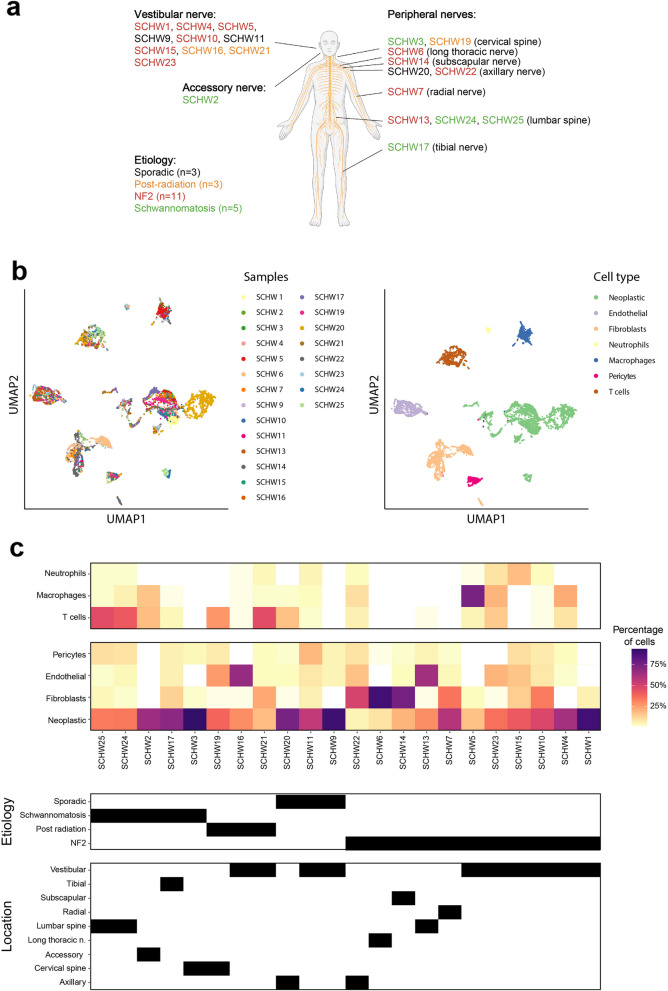
Background: Schwannomas are nerve sheath tumors arising at cranial and peripheral nerves, either sporadically or in patients with a schwannomatosis-predisposition syndrome. There is limited understanding of the transcriptional heterogeneity of schwannomas across genetic backgrounds and anatomic locations.
Methods: Here, we prospectively profile by single-cell full-length transcriptomics tumors from 22 patients with NF2-related schwannomatosis, non-NF2-related schwannomatosis, and sporadic schwannomas, resected from cranial and peripheral nerves. We profiled 11,373 cells (after QC), including neoplastic cells, fibroblasts, T cells, endothelial cells, myeloid cells, and pericytes.
Results: We characterize the intra-tumoral genetic and transcriptional heterogeneity of schwannoma, identifying six distinct transcriptional metaprograms, with gene signatures related to stress, myelin production, antigen presentation, interferon signaling, glycolysis, and extracellular matrix. We demonstrate the robustness of our findings with analysis of an independent cohort.
Conclusions: Overall, our atlas describes the spectrum of gene expression across schwannoma entities at the single-cell level and will serve as an important resource for the community.
Keywords: NF2; Neurofibromatosis type 2; Schwannoma; Schwannomatosis.
© 2025. The Author(s).
Conflict of interest statement
Declarations. Ethics approval and consent to participate: This study received ethics approval from the Institutional Review Board of the Dana-Farber / Harvard Cancer Center (DF/HCC Protocol 10- 417). Written informed consent to participate was obtained from all individuals whose tumor samples were included in this study. The research above described is in conformity with the principles of the Helsinki Declaration. Consent for publication: Not applicable. Competing interests: LNGC [Employment (Takeda - spouse), Stock (Takeda - spouse), Consulting (Oakstone Publishing, Elsevier, BMJ Publishing, Prime Education, Servier Laboratories), Research Funding (Merck - to DFCI)]; JTJ [Employment (Elsevier), Stock (Akeila Bio, Navio Theragnostics, The Doctor Lounge), Consulting (Akeila Bio, Alexion Pharmaceuticals, Magnet Biomedicine, Navio Theragnostics, Recursion Pharmaceuticals, Shepherd Therapeutics, Springwork Therapeutics)]; SRP [Cofounder (NF2 therapeutics), Consulting (Akouos)]; MLS [Cofounder (Immunitas Therapeutics), Stock (Immunitas Therapeutics)]. The remaining authors declare that they do not have any competing interests.
Figures


Similar articles
-
A mosaic pattern of INI1/SMARCB1 protein expression distinguishes Schwannomatosis and NF2-associated peripheral schwannomas from solitary peripheral schwannomas and NF2-associated vestibular schwannomas.Childs Nerv Syst. 2017 Jun;33(6):933-940. doi: 10.1007/s00381-017-3340-2. Epub 2017 Apr 1. Childs Nerv Syst. 2017. PMID: 28365909
-
Clinical features of spinal schwannomas in 65 patients with schwannomatosis compared with 831 with solitary schwannomas and 102 with neurofibromatosis Type 2: a retrospective study at a single institution.J Neurosurg Spine. 2016 Jan;24(1):145-54. doi: 10.3171/2015.3.SPINE141145. Epub 2015 Sep 25. J Neurosurg Spine. 2016. PMID: 26407091
-
A group of schwannomas with interstitial deletions on 22q located outside the NF2 locus shows no detectable mutations in the NF2 gene.Hum Genet. 1999 May;104(5):418-24. doi: 10.1007/s004390050978. Hum Genet. 1999. PMID: 10394935
-
The neurofibromatoses. Part 2: NF2 and schwannomatosis.Rev Neurol Dis. 2009 Summer;6(3):E81-6. Rev Neurol Dis. 2009. PMID: 19898272 Review.
-
[Segmental schwannomatosis in upper-extremity: 5 cases report and literature review].Beijing Da Xue Xue Bao Yi Xue Ban. 2013 Oct 18;45(5):698-703. Beijing Da Xue Xue Bao Yi Xue Ban. 2013. PMID: 24136261 Review. Chinese.
Cited by
-
SMARCB1-related schwannomatosis and other SMARCB1-associated phenotypes: clinical spectrum and molecular pathogenesis.Fam Cancer. 2025 Aug 12;24(3):64. doi: 10.1007/s10689-025-00486-4. Fam Cancer. 2025. PMID: 40794298 Free PMC article. Review.
References
-
- WHO Classification of Tumours Editorial Board. Central nervous system tumors. 5th ed. Lyon: International Agency for Research on Cancer; 2021.
-
- Nonaka Y, Fukushima T, Watanabe K, Friedman AH, Cunningham CD, Zomorodi AR. Surgical management of vestibular schwannomas after failed radiation treatment. Neurosurg Rev. 2016;39:303–12. - PubMed
-
- Frischer JM, Gruber E, Schöffmann V, Ertl A, Höftberger R, Mallouhi A, et al. Long-term outcome after Gamma Knife radiosurgery for acoustic neuroma of all Koos grades: a single-center study. J Neurosurg. 2019;130:388–97. - PubMed
-
- Klijn S, Verheul JB, Beute GN, Leenstra S, Mulder JJS, Kunst HPM, et al. Gamma knife radiosurgery for vestibular schwannomas: evaluation of tumor control and its predictors in a large patient cohort in the Netherlands. J Neurosurg. 2016;124:1619–26. - PubMed
-
- Huang C, Tu H, Chuang C, Chang C, Chou H, Lee M, et al. Gamma knife radiosurgery for large vestibular schwannomas greater than 3 cm in diameter. J Neurosurg. 2018;128:1380–7. - PubMed
MeSH terms
LinkOut - more resources
Full Text Sources
Research Materials
Miscellaneous
