. 2025 Apr 11;23(1):32.
doi: 10.1186/s12959-025-00715-x.

Tissue factor, factor VIII and IX in microvesicle-induced thrombosis and tumor growth of pancreatic cancer

Affiliations

Tissue factor, factor VIII and IX in microvesicle-induced thrombosis and tumor growth of pancreatic cancer

Sheng-Chieh Chou et al. Thromb J. .

Abstract

Background: Tissue factor (TF)-rich cancer microvesicles are correlated with thrombosis risk. Intrinsic coagulation factors are also associated with the risk of thrombosis in cancer patients. This study explored the roles of pancreatic cancer-derived microvesicles and intrinsic factors in thrombogenesis.

Methods: Human pancreatic cancer cell lines rich in TF (AsPC-1-TFhigh, MIAPaCa-2-TFhigh) or poor in TF [AsPC-1-TFKO(knockout) and MIAPaCa-2-TFlow] were generated for microvesicle preparation and injected into coagulation-defective mice. Inferior vena cava (IVC) clots and lung thrombosis were evaluated. Immunodeficient hemophilia A (NSG-HA) mice were orthotopically injected with the cells mentioned above, and the tumor and IVC clot weights were analyzed.

Results: With the injection of TFhigh microvesicles, IVC clots were rarely found in hemophilic mice. The TFlow and TFKO microvesicles resulted in few IVC clots in any mouse. Lung thrombosis was substantially reduced in the hemophilic mice infused with any microvesicle type. In orthotopic tumor models, TFhigh cells grew faster than did TFlow cells. TFhigh tumor-bearing NSG-WT mice had the most enormous IVC clots, whereas NSG-HA mice had no IVC clots.

Conclusion: Pancreatic cancer thrombosis induced by TF-expressing microvesicles strongly depended on FVIII and FIX, while VWF played a minor role. Moreover, TF, but not FVIII, was significantly related to tumor growth.


Conflict of interest statement

Declarations. Competing interests: The authors declare no competing interests.

Figures

Fig. 1
Fig. 1
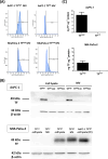
Size distribution of MVs derived from AsPC-1, MIA PaCa-2 cells was conducted using nanoparticle tracking analysis. Representative histograms are shown
Fig. 2
Fig. 2
(A, B) Structure of MVs derived from (A) AsPC-1 (TFhigh) and AsPC-1 (TFKO) and (B) MIA PaCa-2 (TFhigh) and MIA PaCa-2 (TFlow), imaged by transmission electron microscopy at 20,000x and 50,000x magnification. Scale bars: 1 μm and 200 nm, respectively. (C) Western blot analysis of CD81 expression in MVs derived from AsPC-1 and MIA PaCa-2 cells (20 µg/well). α-Tubulin and β-actin were used as internal controls
Fig. 3
Fig. 3
(A) Expression of TF on MVs was determined using an APC-conjugated anti-human TF antibody by flow cytometry, with gating based on an unstained sample (data not shown). The horizontal axis represents TF-positive cells, and the vertical axis represents cell count. (B) Western blot analysis of TF expression in AsPC-1 and MIA PaCa-2 cells and MVs (20 µg/well). AsPC-1 TFhigh cell lysate (PC) was used as positive control of TF expression and β-actin was used as internal control. (C) TF activity was measured using a 1-stage clotting assay
Fig. 4
Fig. 4
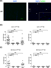
(A) Clot in the IVC model of WT mice injected with MIA PaCa-2 TFhigh MVs, in which the MVs were stained with rabbit anti-human CD81 antibody followed by Alexa Fluor® 488 conjugated goat anti-rabbit antibody and analyzed by immunofluorescence. Nuclei were stained with DAPI. Scale bar, 100 μm. Insert, magnification of the dashed box. Scale bar, 50 μm. The left panel shows staining with DAPI alone as a control, while the right panel displays staining with Anti- human CD81 antibody. (B) After IVC stenosis was achieved, 40 µg of MV from AsPC-1 (TFhigh/TFKO) cells were injected intravenously into the WT (n = 31/12), HA (n = 19/7), HB (n = 13/7) and vWD mice (n = 18/7); (C) After IVC stenosis was achieved, 40 µg of MV from MIA PaCa-2 (TFhigh/ TFlow) cells were injected intravenously into the WT (n = 14/5), HA (n = 8/7), HB (n = 7/9) and vWD mice (n = 11/6). Each dot represents one mouse, with a line indicating the mean. * p ≤ 0.05; ** p ≤ 0.01; *** p ≤ 0.001; **** p ≤ 0.0001; NS, non-significant
Fig. 5
Fig. 5
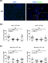
(A) Lung tissue sections from the IVC model of WT mice injected with MIA PaCa-2 TFhigh MVs, in which the MVs were stained with rabbit anti-human CD81 antibody followed by Alexa Fluor® 488 conjugated goat anti-rabbit antibody and analyzed by immunofluorescence. Nuclei were stained with DAPI. The left panel shows staining with DAPI alone as a control, while the right panel displays staining with Anti- human CD81 antibody. Scale bar, 100 μm. Insert, magnification of the dashed box. Scale bar, 50 μm. (B, C) Lung tissue sections stained with H&E were examined at 400x magnification. The number of thrombi within vessels was counted from twenty randomly selected observation fields, and the results are summarized as the representative measure of lung thrombosis. Each dot represents one mouse, with n = 5–9 per group. Lines in dot plots represent the mean. * p ≤ 0.05; ** p ≤ 0.01; *** p ≤ 0.001; **** p ≤ 0.0001; NS, not significant
Fig. 6
Fig. 6
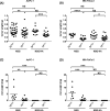
TF supports tumor growth and IVC clots. 1 × 106 AsPC-1 (TFhigh/TFKO) and MIA PaCa-2 (TFhigh/TFlow) cells were orthotopically injected into the pancreas of NSG and NSG-HA mice. After 5 weeks, IVC flow was completely blocked for 1 h, and the mice were sacrificed to measure (A, B) tumor weight and (C, D) clot weight. * Each dot represents one mouse, with n = 6–32 per group. Lines in dot plots represent the mean. p ≤ 0.05; ** p ≤ 0.01; *** p ≤ 0.001; **** p ≤ 0.0001; NS, non-significant

References

    1. Khorana AA, Francis CW, Culakova E, Kuderer NM, Lyman GH. Thromboembolism is a leading cause of death in cancer patients receiving outpatient chemotherapy. J Thromb Haemost. 2007;5:632–4. - PubMed
    1. Mulder FI, Horvath-Puho E, van Es N, van Laarhoven HWM, Pedersen L, Moik F, Ay C, Buller HR, Sorensen HT. Venous thromboembolism in cancer patients: a population-based cohort study. Blood. 2021;137:1959–69. - PubMed
    1. Khorana AA, Mackman N, Falanga A, Pabinger I, Noble S, Ageno W, Moik F, Lee AYY. Cancer-associated venous thromboembolism. Nat Rev Dis Primers. 2022;8:11. - PubMed
    1. Khorana AA, Kuderer NM, Culakova E, Lyman GH, Francis CW. Development and validation of a predictive model for chemotherapy-associated thrombosis. Blood. 2008;111:4902–7. - PMC - PubMed
    1. Horsted F, West J, Grainge MJ. Risk of venous thromboembolism in patients with cancer: a systematic review and meta-analysis. PLoS Med. 2012;9:e1001275. - PMC - PubMed

LinkOut - more resources