. 2025 Mar 25;14(7):2246.
doi: 10.3390/jcm14072246.

A New Approach for Reconstruction of Severe Horizontal Atrophy of the Posterior Mandible Using "The Honeycomb Technique": A 10-14 Year Follow-Up Retrospective Study

Affiliations

A New Approach for Reconstruction of Severe Horizontal Atrophy of the Posterior Mandible Using "The Honeycomb Technique": A 10-14 Year Follow-Up Retrospective Study

Fares Kablan. J Clin Med. .

Abstract

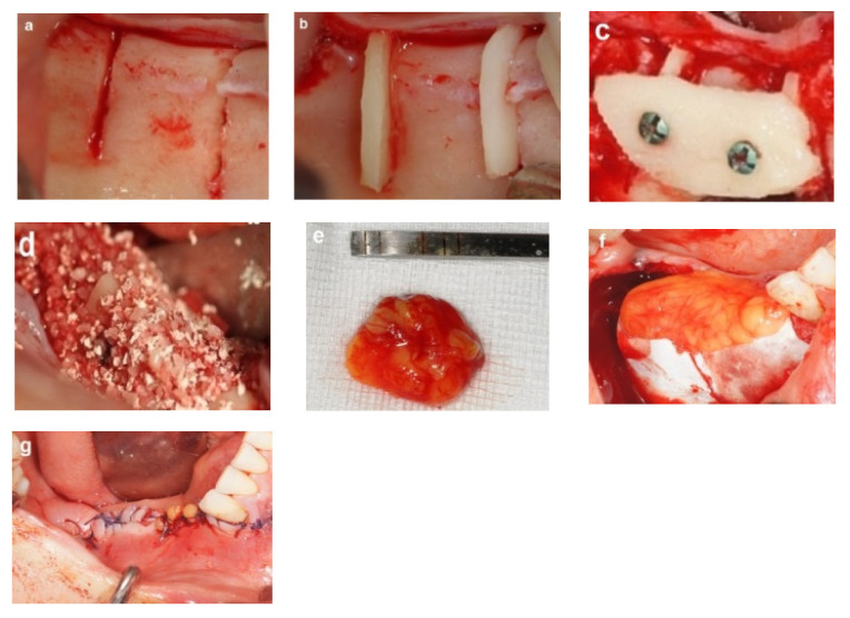
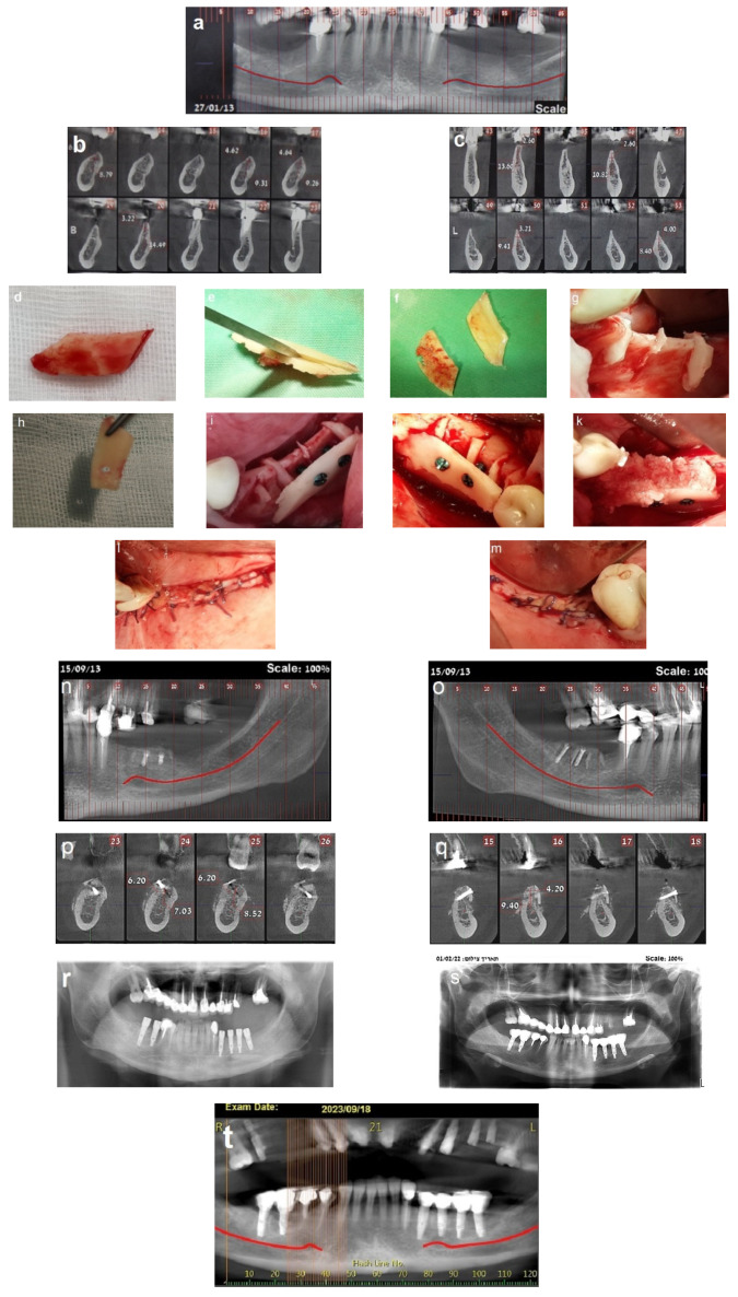
Background: Autogenous bone grafting has long been the standard for augmenting bone prior to implant placement in atrophic ridges. However, innovative techniques are continually sought to enhance outcomes. This study introduces the honeycomb technique for horizontal bone augmentation in edentulous posterior mandibular ridges, presenting the methodology and long-term follow-up results of this novel approach. Methods: This study includes healthy patients with moderate to severe horizontal atrophy in posterior mandibular regions who underwent bone augmentation using the honeycomb technique and were followed up for a period of 10 to 14 years. The patients had orthoradiographs immediately post-surgery and underwent regular clinical and radiographic evaluations. Computed tomography at four months assessed the bone gain, followed by reentry for implant insertion and evaluation of the bone volume and quality. Fixed prosthesis-supported dental implants were placed four months post-insertion. The survival and success of the dental implants were evaluated based on the acceptable clinical and radiographic criteria. Results: A cohort of 23 patients (17 women, 6 men, mean age 47 years) underwent bone augmentation at 39 sites, with follow-up ranging from 10 to 14 years. The procedure demonstrated a 95-100% success rate with minimal morbidity and horizontal bone gain averaging 3-8 mm. Partial graft exposure occurred in two cases but was successfully managed without compromising augmentation. A total of 103 implants were placed in the augmented sites in 37 sites. The long-term survival of the dental implants was confirmed based on clinical and radiographic evaluation, with minimal marginal bone loss observed during the extended follow-up period. Conclusions: The honeycomb technique proves effective in horizontal bone augmentation of atrophic ridges in posterior mandibular defects. The satisfactory long-term outcomes validate its potential as a valuable addition to bone augmentation strategies preceding implant placement.

Keywords: allogenic bone substitute; autogenic bone grafts; bone augmentation; mandibular atrophy; space maintenance.


Conflict of interest statement

The author declares no conflicts of interest.

Figures

Figure 1
Figure 1
(a) Bilateral posterior mandibular edentulism. (bd) Computed tomography showing severe horizontal atrophy with knife-edge residual ridges.
Figure 2
Figure 2
(a) The donor site. (b) The bone block harvested from the retromolar donor site. (ce) The harvested bone block and its subsequent splitting, resulting in thin bone blocks and cortical wedges.
Figure 3
Figure 3
The bone augmentation procedure. (a) Grooves created using high-speed bur at the recipient site. (b) Insertion and tapping of the cortical bone wedges into the grooves. (c) Positioning a thin bone plate over the bone wedges and securing it with screws, forming bone compartments resembling a honeycomb. (d) Filling the bone compartments with allogenic particulate bone graft to achieve the desired bone graft volume. (e) Free buccal fat tissue graft (BFFG). (f) Resorbable membrane and BFFG covering the augmented bone. (g) Double-layer tension-free closure of the recipient site.
Figure 4
Figure 4
Reentry at 4 months. (ac) CBCT post-op demonstrating significant horizontal bone gain. (d) Left-side view, showing excellent integration of the bone blocks and the width of the new bone demonstrated by the length of the removed fixation screws (e). (f,g) Placement of three implants on the left side. (h) New bone volume on the right side. (i) Placement of three implants on the right side.
Figure 5
Figure 5
Follow-up. (a,b) Ten-year follow-up post-loading, showing the implant at the recipient site with no marginal bone loss.
Figure 6
Figure 6
(a) Bilateral posterior mandibular edentulism. (b,c) Computed tomography showing severe horizontal atrophy with knife-edge residual ridges. (d) The bone block harvested from the retromolar donor site. (e,f) The harvested bone block and its subsequent splitting, resulting in thin bone blocks and cortical wedges. (g) The bone augmentation procedure: grooves were created using high-speed at the recipient site, followed by the insertion and tapping of the cortical bone wedges into the grooves. (h,i) A thin bone plate is positioned over the bone wedges and secured with screws, creating bone compartments resembling a honeycomb. (j) Right side. (k) The bone compartments are filled with allogenic particulate bone graft to achieve the desired graft volume on the right side. (l,m) Double-layer, tension-free closure of the recipient sites performed using BFFGs. (nq) CBCT scan, four months postoperatively show significant bilateral horizontal bone gain. (r) Two implants were placed on the right side and four implants on the left side. (s) Panoramic view at 8-year follow-up. (tv) CBCT scans taken 10 years after implant placement reveal stable augmented bone levels. (w) Eleven-year follow-up periapical x-ray of the left side, demonstrating no significant marginal bone loss. (x) 3 mm of marginal bone loss observed at the mesial implant. The yellow arrows show the cortical wedges that remain visible at the recipient sites.
Figure 6
Figure 6
(a) Bilateral posterior mandibular edentulism. (b,c) Computed tomography showing severe horizontal atrophy with knife-edge residual ridges. (d) The bone block harvested from the retromolar donor site. (e,f) The harvested bone block and its subsequent splitting, resulting in thin bone blocks and cortical wedges. (g) The bone augmentation procedure: grooves were created using high-speed at the recipient site, followed by the insertion and tapping of the cortical bone wedges into the grooves. (h,i) A thin bone plate is positioned over the bone wedges and secured with screws, creating bone compartments resembling a honeycomb. (j) Right side. (k) The bone compartments are filled with allogenic particulate bone graft to achieve the desired graft volume on the right side. (l,m) Double-layer, tension-free closure of the recipient sites performed using BFFGs. (nq) CBCT scan, four months postoperatively show significant bilateral horizontal bone gain. (r) Two implants were placed on the right side and four implants on the left side. (s) Panoramic view at 8-year follow-up. (tv) CBCT scans taken 10 years after implant placement reveal stable augmented bone levels. (w) Eleven-year follow-up periapical x-ray of the left side, demonstrating no significant marginal bone loss. (x) 3 mm of marginal bone loss observed at the mesial implant. The yellow arrows show the cortical wedges that remain visible at the recipient sites.
Figure 7
Figure 7
(ac) CBCT scan images reveal severe horizontal atrophy in the molar regions bilaterally. (df) CBCT scan images taken after honeycomb bone grafting demonstrate significant bone gain. (g); Implants, (h,i) images taken 13-year follow-up after implant loading.
Figure 7
Figure 7
(ac) CBCT scan images reveal severe horizontal atrophy in the molar regions bilaterally. (df) CBCT scan images taken after honeycomb bone grafting demonstrate significant bone gain. (g); Implants, (h,i) images taken 13-year follow-up after implant loading.
Figure 8
Figure 8
(a) Patient No. 1; follow-up 14 years. (b) Patient No. 5; follow-up 14 years.
Figure 9
Figure 9
(a,b) Ten-year follow-up periapical x-rays reveal severe bone loss involving the distal implant on the left side, along with 3 mm of marginal bone loss at the middle implant on the right side. (ce) The images demonstrate stable bone grafts despite the severe marginal bone loss at the distal implant on the left side.
Figure 10
Figure 10
Panoramic x-ray taken 13 years after implant loading reveals severe marginal bone loss involving the distal implant, along with a fracture of the mesial implant hex.
Figure 11
Figure 11
(a) A panoramic x-ray taken 13 years after implant loading demonstrates stable outcomes across five implants, though the middle implant on the right side exhibits 3 mm marginal bone loss. (b) A panoramic x-ray taken 11.5 years after implant loading shows stable outcomes for five implants, with 3 mm marginal bone loss affecting the distal implant on the right side.
Chart 1
Chart 1
The bar chart illustrates the percentage of surviving implants (97%), failed implants, successful implants (93.2%), and implants with marginal bone loss of 30%.
Chart 2
Chart 2
Kaplan–Meier survival analysis for dental implants over a follow-up period of 10 to 14 years.

Similar articles

References

    1. Wang Y., Baumer D., Ozga A.N., Korner G., Baumer A. Patient satisfaction and oral health-related quality of life 10 years after implant placement. BMC Oral Health. 2021;21:30. doi: 10.1186/s12903-020-01381-3. - DOI - PMC - PubMed
    1. Babu R.S.A., Ogle O. Tissue response: Biomaterials, dental implants, and compromised osseous tissue. Dent. Clin. N. Am. 2015;59:305–315. doi: 10.1016/j.cden.2014.10.010. - DOI - PubMed
    1. Tan W.L., Wong T.L., Wong M.C., Lang N.P. A systematic review of post-extraction alveolar hard and soft tissue dimensional changes in humans. Clin. Oral Implant. Res. 2012;23((Suppl. S5)):1–21. doi: 10.1111/j.1600-0501.2011.02375.x. - DOI - PubMed
    1. Felice P., Zucchelli G., Cannizzaro G., Barausse C., Diazzi M., Trullenque-Eriksson A., Esposito M. Immediate, immediate-delayed (6 weeks) and delayed (4 months) post-extractive single implants: 4-month post-loading data from a randomized controlled trial. Eur. J. Oral Implantol. 2016;9:233–247. - PubMed
    1. Alenazi A., Alotaibi A.A., Aljaeide Y.M., Alqhtani N.R. The need for socket preservation: A systematic review. J. Med. Life. 2022;15:309–312. doi: 10.25122/jml-2021-0308. - DOI - PMC - PubMed

LinkOut - more resources