

Advances in Skin Ultrasonography for Malignant and Benign Tumors of the Head and Neck: Current Insights and Future Directions
- PMID: 40217748
- PMCID: PMC11989985
- DOI: 10.3390/jcm14072298
Advances in Skin Ultrasonography for Malignant and Benign Tumors of the Head and Neck: Current Insights and Future Directions
Abstract
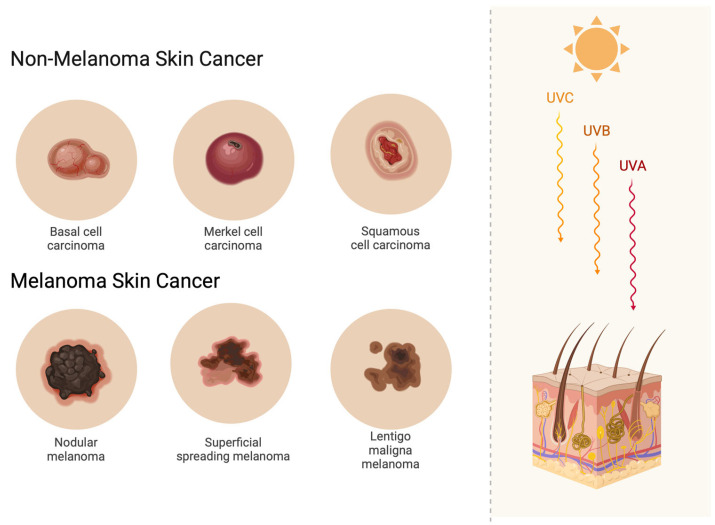
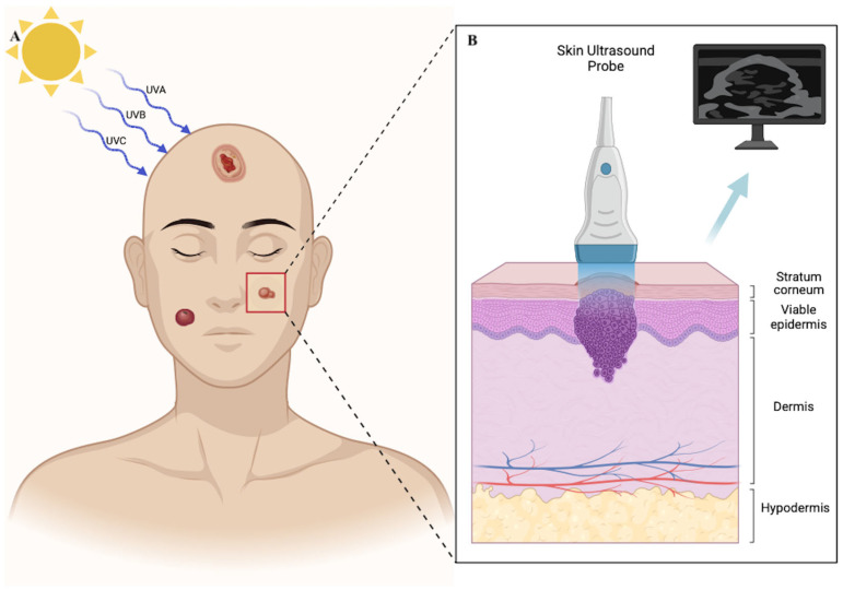
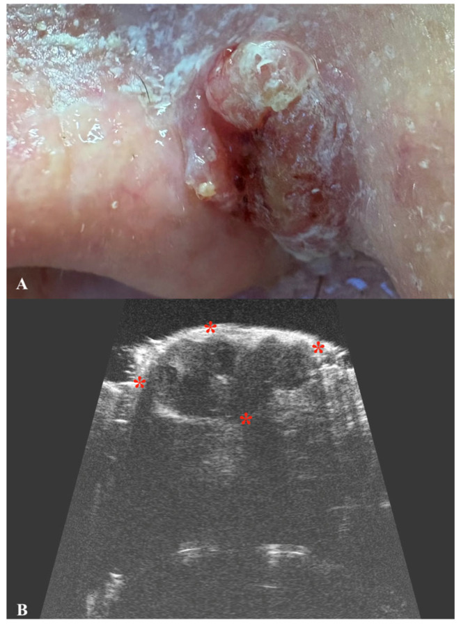
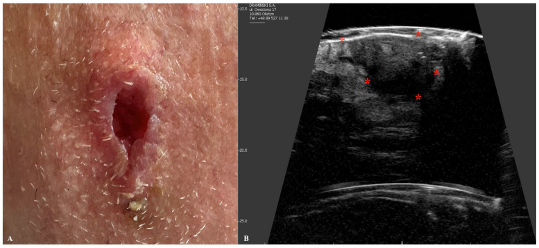
Ultrasound imaging has become an indispensable diagnostic tool across various medical fields. In recent years, there has been growing interest in the use of ultrasonography for the evaluation of skin lesions. However, scientific reports detailing the precise role of ultrasound in determining the morphology of malignant skin tumors still remain limited. Malignant skin lesions, particularly in the head and neck region-their most common location-pose significant challenges due to the complex anatomy of these areas. The primary treatment for non-melanoma skin cancers, including basal cell carcinoma (BCC) and squamous cell carcinoma (SCC), is surgical excision. Mohs micrographic surgery is considered the gold standard due to its tissue-sparing approach and high cure rates. However, it is a time-consuming and resource-intensive procedure that is not always widely accessible. In contrast, standard surgical excision, while more widely available, often results in incomplete tumor removal, necessitating subsequent surgical radicalization or the use of adjuvant therapies. Routine ultrasound evaluation of both benign and malignant skin lesions could enhance early detection and facilitate timely treatment. However, the current body of evidence for the usage of skin ultrasound in presurgical evaluation is poor and lacks standardization. Given these challenges, in this review, we aim to highlight the potential value of preoperative skin ultrasonography in accurately assessing benign and malignant skin lesion dimensions and morphology.
Keywords: Mohs surgery; basal cell carcinoma; high-frequency ultrasonography; melanoma; squamous cell carcinoma.
Conflict of interest statement
The authors declare no conflicts of interest.
Figures

Similar articles
-
Diagnostic accuracy of autofluorescence-Raman microspectroscopy for surgical margin assessment during Mohs micrographic surgery of basal cell carcinoma.Br J Dermatol. 2024 Aug 14;191(3):428-436. doi: 10.1093/bjd/ljae196. Br J Dermatol. 2024. PMID: 38736216
-
A relative value unit-based cost comparison of treatment modalities for nonmelanoma skin cancer: effect of the loss of the Mohs multiple surgery reduction exemption.J Am Acad Dermatol. 2009 Jul;61(1):96-103. doi: 10.1016/j.jaad.2008.07.047. J Am Acad Dermatol. 2009. PMID: 19539843
-
Surgical excision of non-melanoma skin cancer: no end in site?Br J Oral Maxillofac Surg. 2021 Dec;59(10):1264-1269. doi: 10.1016/j.bjoms.2021.05.018. Epub 2021 Jun 5. Br J Oral Maxillofac Surg. 2021. PMID: 34275678
-
Who should have Mohs micrographic surgery?Curr Opin Otolaryngol Head Neck Surg. 2010 Aug;18(4):283-9. doi: 10.1097/MOO.0b013e32833b6f19. Curr Opin Otolaryngol Head Neck Surg. 2010. PMID: 20613530 Review.
-
Mohs micrographic surgery: a review of indications, technique, outcomes, and considerations.An Bras Dermatol. 2021 May-Jun;96(3):263-277. doi: 10.1016/j.abd.2020.10.004. Epub 2021 Mar 24. An Bras Dermatol. 2021. PMID: 33849752 Free PMC article. Review.
Cited by
-
A Novel Radiology-Adapted Logistic Model for Non-Invasive Risk Stratification of Pigmented Superficial Skin Lesions: A Methodological Pilot Study.Diagnostics (Basel). 2025 Jul 30;15(15):1921. doi: 10.3390/diagnostics15151921. Diagnostics (Basel). 2025. PMID: 40804885 Free PMC article.
References
-
- Situm M., Buljan M., Bulat V., Lugović Mihić L., Bolanca Z., Simić D. The role of UV radiation in the development of basal cell carcinoma. Coll. Antropol. 2008;32((Suppl. S2)):167–170. - PubMed
Publication types
Grants and funding
LinkOut - more resources
Full Text Sources
Research Materials
