

Entire Papilla Preservation Technique with Enamel Matrix Proteins and Allogenic Bone Substitutes for the Treatment of Isolated Intrabony Defects: A 3-Year Follow-Up of a Prospective Case Series
- PMID: 40217825
- PMCID: PMC11989921
- DOI: 10.3390/jcm14072374
Entire Papilla Preservation Technique with Enamel Matrix Proteins and Allogenic Bone Substitutes for the Treatment of Isolated Intrabony Defects: A 3-Year Follow-Up of a Prospective Case Series
Abstract
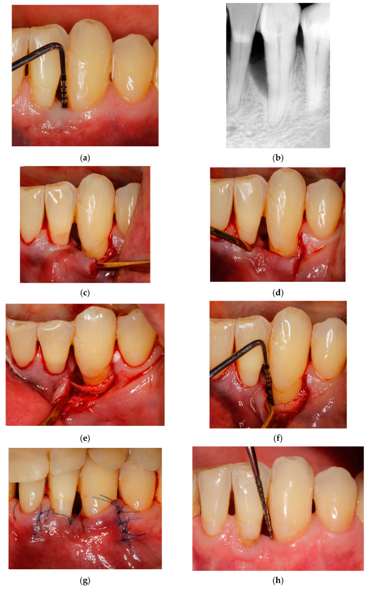
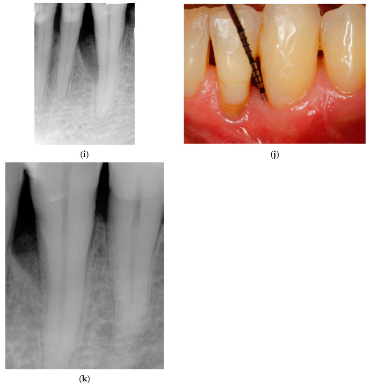
Background: This study aimed to assess the effectiveness of a modified entire papilla preservation technique (MEPPT) for treating isolated intrabony defects in patients with stage III periodontitis. Material and Methods: Fifteen patients with 15 interdental intrabony defects were treated with a MEPPT using enamel matrix derivative and allogenic bone. Their probing pocket depth (PPD), clinical attachment level (CAL), gingival recession (GR), keratinized tissue width (KTW), defect depth (DD), full-mouth plaque score (FMPS), full mouth bleeding score (FMBS), radiographic images (radiographic angles, BF and LDF) and intrasurgical parameters were assessed at baseline and 3 years postsurgery. Standardized measurements were taken to evaluate the defect characteristics and treatment outcomes. Results: At 3 years, significant improvements from baseline were maintained. Probing pocket depth (PPD) decreased from 7.03 ± 1.61 mm to 3.33 ± 0.89 mm (p < 0.0001), clinical attachment level (CAL) improved to 3.08 ± 1.16 mm (p < 0.001) and defect depth (DD) decreased from 4.59 ± 1.24 mm to 0.38 ± 0.31 mm (p < 0.001). The changes in gingival recession and keratinized tissue were not statistically significant. The results demonstrate sustained clinical stability over a 3-year period. Conclusions: Within the limitations of this study, the findings suggest that the modified entire papilla preservation technique (MEPPT) in conjunction with enamel matrix proteins and allogenic bone grafting is an effective approach for the treatment of intrabony defects, leading to statistically significant and sustained clinical improvements over a 3-year period. The study protocol was registered in ClinicalTrials.gov ID NCT05029089.
Keywords: allogenic bone; enamel matrix protein; intrabony defect; modified entire papilla preservation technique; periodontitis.
Conflict of interest statement
The authors declare no conflicts of interest.
Figures

References
-
- Lang N.P., Salvi G.E., Sculean A. Nonsurgical Therapy for Teeth and Implants—When and Why? Periodontology 2000. 2019;79:15–21. - PubMed
-
- Tomasi C., Leyland A.H., Wennstrom J.L. Factors Influencing the Outcome of Non-Surgical Periodontal Treatment: A Multilevel Approach. J. Clin. Periodontol. 2007;34:682–690. - PubMed
-
- Cortellini P., Tonetti M.S. Clinical concepts for regenerative therapy in intrabony defects. Periodontology 2000. 2015;68:282–307. - PubMed
Publication types
Associated data
LinkOut - more resources
Full Text Sources
Medical
Miscellaneous
