Review
. 2025 Mar 31;14(7):2396.
doi: 10.3390/jcm14072396.

Techniques of Deformity Correction in Adolescent Idiopathic Scoliosis-A Narrative Review of the Existing Literature

Affiliations
Review

Techniques of Deformity Correction in Adolescent Idiopathic Scoliosis-A Narrative Review of the Existing Literature

Aakash Jain et al. J Clin Med. .

Abstract

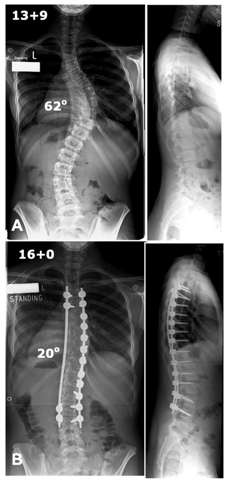
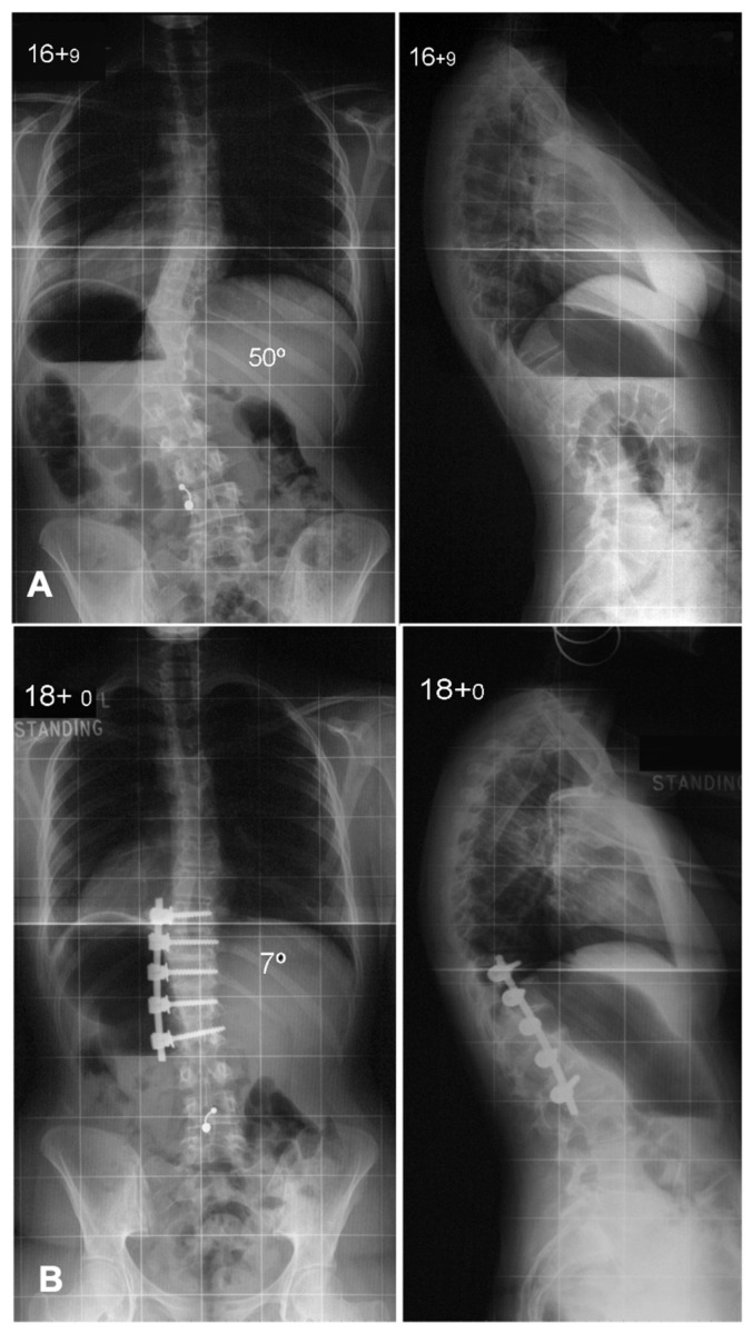
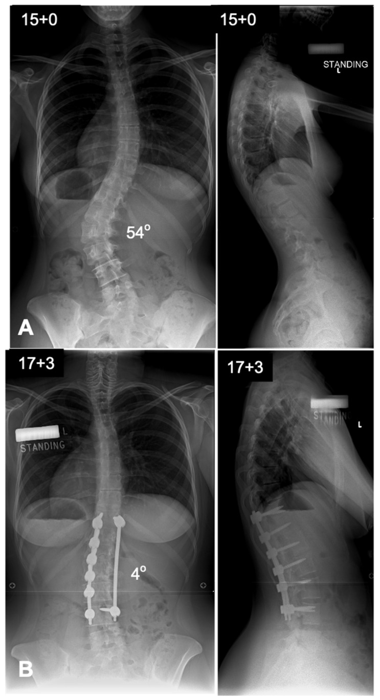
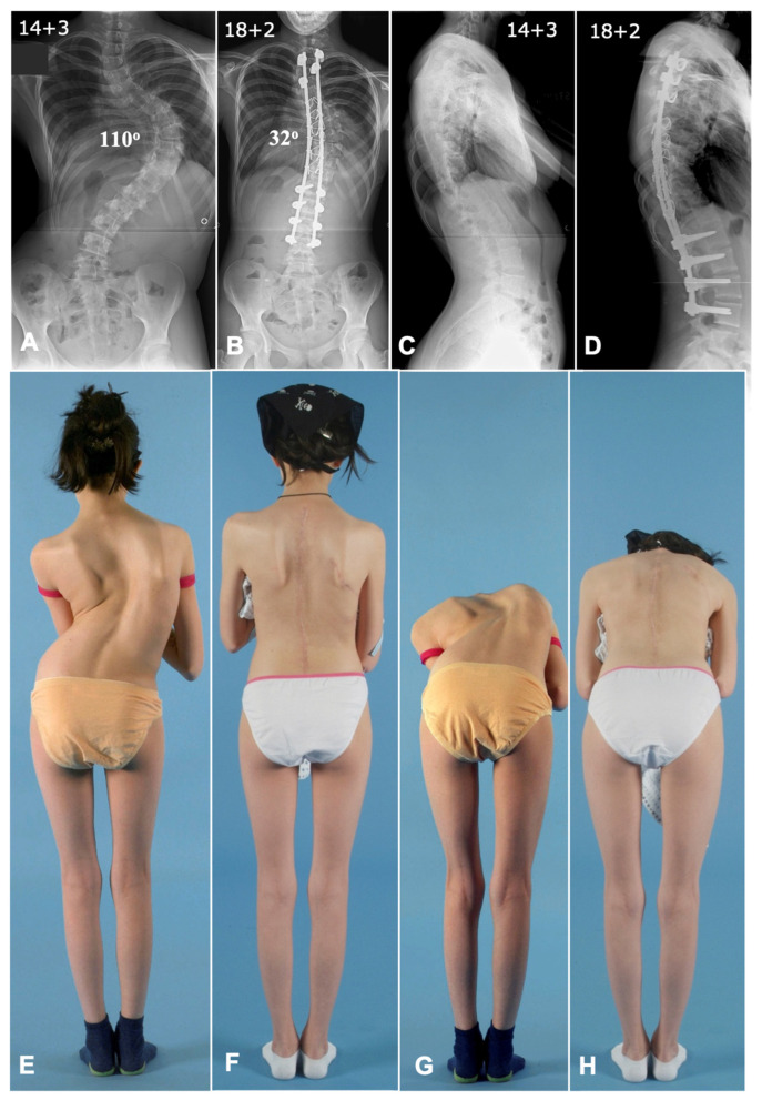
Surgical management of adolescent idiopathic scoliosis [AIS] is a complex undertaking with the primary goals to correct the deformity, maintain sagittal balance, preserve pulmonary function, maximize postoperative function, and improve or at least not harm the function of the lumbar spine. The evolution of surgical techniques for AIS has been remarkable, transitioning from rudimentary methods of spinal correction to highly refined, biomechanically sound procedures. Modern techniques incorporate advanced three-dimensional correction strategies, often leveraging pedicle screw constructs, which provide superior rotational control of the vertebral column. A number of surgical techniques have been described in the literature, each having its own pros and cons. This narrative review provides a detailed analysis of the contemporary surgical techniques used in the treatment of patients with AIS.

Keywords: adolescent idiopathic scoliosis; deformity surgery; surgical techniques.


Conflict of interest statement

The authors declare no conflicts of interest.

Figures

Figure 1
Figure 1
Schwab anatomical spinal osteotomy classification [19].
Figure 2
Figure 2
Deformity correction using a typical concave side rod derotation maneuver. (A) Preoperative antero-posterior and lateral radiographs showing a thoracic scoliosis with a Cobb angle of 48 degrees. (B) Postoperative radiographs showing good deformity correction with coronal and sagittal balance.
Figure 3
Figure 3
Deformity correction using double-rod technique. (A) Preoperative antero-posterior and lateral radiographs showing a thoracic scoliosis with a Cobb angle of 80 degrees. (B) Postoperative radiographs showing good deformity correction with coronal and sagittal balance.
Figure 4
Figure 4
MRI cut sections at (A) D8 and (B) L1 showing relatively safer convex side pedicles (arrows) with the spinal cord lying at a distance from the convex pedicle when compared to its concave counterpart.
Figure 5
Figure 5
Deformity correction using convex rod technique. (A) Preoperative antero-posterior and lateral radiographs showing a thoracic scoliosis with a Cobb angle of 62 degrees. (B) Postoperative radiographs showing good deformity correction with coronal and sagittal balance.
Figure 6
Figure 6
Deformity correction using anterior spinal fusion in a Lenke 5 curve. (A) Preoperative antero-posterior and lateral radiographs showing a thoraco-lumbar scoliosis with a Cobb angle of 50 degrees. (B) Postoperative radiographs showing good deformity correction with coronal and sagittal balance. Anterior spinal fusion allows good deformity correction with shorter fusion constructs.
Figure 7
Figure 7
Deformity correction using posterior spinal fusion in a Lenke 5 curve. (A) Preoperative antero-posterior and lateral radiographs showing a thoraco-lumbar scoliosis with a Cobb angle of 54 degrees. (B) Postoperative radiographs showing good deformity correction with coronal and sagittal balance. Correction in posterior spinal fusion in thoraco-lumbar and lumbar curves is achieved from the convex rod.
Figure 8
Figure 8
Deformity correction using a combined anterior and posterior approach in a severe rigid thoracic scoliosis case. (A,C) Preoperative antero-posterior and lateral radiographs showing a thoracic scoliosis with a Cobb angle of 110 degrees. (B,D) Postoperative radiographs showing good deformity correction with a Cobb angle of 32 degrees with well-maintained coronal and sagittal balance.(E,G) Preoperative clinical photographs showing a severe deformity and rib hump. (F,H) Postoperative clinical photographs showing a good correction of deformity and a well-maintained balance. Note the correction of the convex rib hump by anterior thoracoplasty.

Similar articles

References

    1. Stokes I.A., Bigalow L.C., Moreland M.S. Three-Dimensional Spinal Curvature in Idiopathic Scoliosis. J. Orthop. Res. 1987;5:102–113. doi: 10.1002/jor.1100050113. - DOI - PubMed
    1. Choudhry M.N., Ahmad Z., Verma R. Adolescent Idiopathic Scoliosis. Open Orthop. J. 2016;10:143–154. doi: 10.2174/1874325001610010143. - DOI - PMC - PubMed
    1. Konieczny M.R., Senyurt H., Krauspe R. Epidemiology of Adolescent Idiopathic Scoliosis. J. Child. Orthop. 2013;7:3–9. doi: 10.1007/s11832-012-0457-4. - DOI - PMC - PubMed
    1. Van Hooff M.L., Te Hennepe N., De Kleuver M. Pulmonary Function in Patients with Spinal Deformity: Have We Been Ignorant? Acta Orthop. 2020;91:503–505. doi: 10.1080/17453674.2020.1786267. - DOI - PMC - PubMed
    1. Sanders A.E., Andras L.M., Iantorno S.E., Hamilton A., Choi P.D., Skaggs D.L. Clinically Significant Psychological and Emotional Distress in 32% of Adolescent Idiopathic Scoliosis Patients*. Spine Deform. 2018;6:435–440. doi: 10.1016/j.jspd.2017.12.014. - DOI - PubMed

LinkOut - more resources