. 2025 Mar 31;14(7):2398.
doi: 10.3390/jcm14072398.

Diagnostic and Therapeutic Approaches for Spinal Subarachnoid Hemorrhage Due to Spinal Aneurysms and Other Etiologies

Affiliations

Diagnostic and Therapeutic Approaches for Spinal Subarachnoid Hemorrhage Due to Spinal Aneurysms and Other Etiologies

Biyan Nathanael Harapan et al. J Clin Med. .

Abstract

Background: Spinal subarachnoid hemorrhage (sSAH) is a very rare disease. Detailed information about the natural course, pathogenesis, radiological manifestation, and therapeutic management is lacking. This study aimed to analyze patients diagnosed with sSAH, focusing on the origin, management strategies, and therapeutic approaches to sSAH. Methods: The study included a cohort of patients admitted to the Department of Neurosurgery, LMU University Hospital, LMU Munich, between January 2021 and December 2024 with a confirmed diagnosis of spinal subarachnoid hemorrhage and, among other things, spinal aneurysms. Data on the included patients were recorded with emphasis on demographics, radiological examination (CT, MRI, and DSA), aneurysm-specific characteristics, and clinical outcome. Results: The study included six patients diagnosed with spinal subarachnoid hemorrhage via multimodal imaging. The etiology of sSAH was identified in all cases, encompassing spinal aneurysms in three patients, anticoagulation therapy in two cases, and bony microspurs in one case, with management strategies tailored as either conservative (monitoring and imaging) or surgical (aneurysm resection, arterial feeder coagulation, or evacuation of intraspinal bleeding). No major adverse events were observed, and all the patients demonstrated neurological improvement or exhibited only mild-to-moderate disability during follow-up. Conclusions: Spinal subarachnoid hemorrhage can be due to a ruptured spinal aneurysm, but in some cases, other underlying causes should be considered as the source of the hemorrhage. Given the scarcity of literature on this condition, it is crucial to identify the correct diagnosis and implement a patient-tailored therapeutic approach.

Keywords: SAH; clipping; neurosurgery; spinal aneurysm; spinal subarachnoid hemorrhage; subarachnoid hemorrhage; vascular disorder; vascular pathology.


Conflict of interest statement

The authors declare no conflicts of interest.

Figures

Figure 1
Figure 1
High-resolution contrast-enhanced MR angiography (ceMRA extreme) demonstrated a probably partially thrombosed spinal aneurysm (arrows) perimedullary on the left with a diameter of 4 mm at the T1 vertebral level ((AC): sagittal, coronal, and transversal planes). DSA in the anteroposterior plane confirmed the partially thrombosed spinal aneurysm at the T1 vertebral level (circle), arising from the radiculomedullary T3/4 artery on the left (arrows) (D). Follow-up DSA was performed after 3 months, visualizing the same arterial supply (left T3/4, arrows) without evidence of residual aneurysm perfusion consistent with complete (spontaneous) aneurysm occlusion (E).
Figure 2
Figure 2
Noncontrast head CT in the transversal plane showing minor subarachnoid hemorrhage, primarily in the right temporoparietal region (A), and distinct subdural hemorrhage in the posterior fossa, particularly involving the craniocervical junction (B,C). High-resolution contrast-enhanced MR angiography (ceMRA extreme) in the sagittal (D) and transversal (E) planes, showing a suspected perimedullary aneurysm dorsolaterally on the left at the T2 level (arrows), measuring approximately 3 mm in diameter. Intraoperative visualization of the aneurysm ((F), arrow). Post-resection view following coagulation of the feeding artery, showing no residual aneurysm (G).
Figure 3
Figure 3
MRI showing a 4 mm intramedullary arterial aneurysm (arrows) with homogeneous contrast enhancement at the C2 vertebral base level and associated spinal cord edema/malacia from C2 to C4, likely post-hemorrhagic in origin. (A) Sagittal T2-weighted sequence; (BD) arterial-phase contrast-enhanced MR angiography in the sagittal, coronal, and transversal planes.
Figure 4
Figure 4
DSA in the anteroposterior plane showing the intramedullary arterial aneurysm at the C2 level, associated with an arteriovenous (AV) shunt originating from an arterial feeder of the left vertebral artery at the C3/4 level, consistent with a perimedullary AV fistula (A). Flat-panel CT angiography with volume rendering technique (VRT) reconstruction clearly shows the AV fistula morphology, including the arterial feeder (bottom arrow), the intramedullary aneurysm (middle arrow), and the draining shunt vein (top arrow) (B). Compared to preoperative DSA (C), no residual aneurysm or shunt vein was observed postoperatively, confirming complete occlusion of the AV fistula (D).
Figure 5
Figure 5
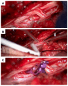
Intraoperative images demonstrating arterial feeder management. Suspected arterial feeder was identified laterally (A). Due to its intramedullary location, the aneurysm was not directly visualized to minimize perioperative morbidity. The use of a micro-Doppler probe allowed identification of the aneurysm (B). Temporary clipping of the feeder was performed (C), followed by intraoperative monitoring (IOM), which remained stable. Doppler assessment indicated no remaining signal, allowing for safe coagulation and transection of the arterial feeder.
Figure 6
Figure 6
Case 4: imaging series illustrating suspected thoracic sub-/intradural bleeding and contrast-enhanced findings potentially indicative of active hemorrhage. CT myelography ((A,B): transversal and sagittal planes) showed evidence of an extensive dorsal sub-/intradural hemorrhage of the thoracic spine (arrows). Transversal (C) and sagittal (D) planes of a body CT scan in the portal-venous phase revealed a streak-like spinal contrast enhancement at the T11 vertebral level, located most likely perimedullarily and dorsolaterally on the right (arrows). This finding, considered a potential “spot sign,” suggested possible active spinal bleeding, likely related to apixaban therapy. Nine days later, a follow-up CT scan in the portal-venous phase ((E,F): transversal and sagittal planes) did not reveal signs of active spinal bleeding any longer, as the suspicious contrast-enhanced lesion at the T11 level had disappeared (circled region). Note: an initial thoracic and lumbar DSA did not reveal any vascular pathology (not shown). Case 5: transversal planes at the levels of C7 (G) and T3 (H), alongside a sagittal view (I) on an MRI T2-weighted sequence, demonstrate a space-occupying intradural hematoma extending from C6 to T3 on the left side. The hematoma exhibited a crescent-shaped configuration, displacing and compressing the spinal cord to the right both ventrally (G) and dorsally (H). Additionally, subarachnoid hemorrhage components extended caudally, and a long-segment myelopathy was evident, spanning from C3 to T6.
Figure 7
Figure 7
Spinal MRI of acute and subacute epi-/subdural hemorrhage with a suspected thrombosed perimedullary aneurysm at the T1/2 level. Initial MRI showed an acute epi-/subdural hemorrhage extending ventrally from levels C1 to T4, with maximal prominence at level T1 (arrows; T2-weighted sequence in the sagittal (A) and transversal planes (B)). The follow-up MRI five days later ((C): sagittal T2-weighted sequence; (D): transversal contrast-enhanced T1-weighted sequence) revealed ongoing (subacute) epi-/subdural bleeding from levels C7 to T3 ventrally (the arrow in (D)) and a newly suspected thrombosed aneurysm with slight wall enhancement at the T1/2 level, located perimedullarily and ventrolaterally on the right (circles in (C,D)). Follow-up MRI two weeks after the initial scan ((E,F): sagittal and transversal T2 -weighted sequence) revealed regression of the hematoma, with no remaining evidence of the aneurysm, suggesting probably complete occlusion (circled area). Spinal CT demonstrated a bony spur (circled area) at the dorsal margin of the intervertebral T1/2 level ((G,H): sagittal and transversal planes). Note: two DSAs were performed, with no vascular pathology detected.

Similar articles

References

    1. Santana-Ramirez A., Farias-Serratos F., Garzon-Muvdi T., Quinones-Hinojosa A. A giant spinal arterial aneurysm in a child presenting as quadriparesis. BMJ Case Rep. 2013;2013:bcr2013200298. doi: 10.1136/bcr-2013-200298. - DOI - PMC - PubMed
    1. Donauer E., Aguilar Perez M., Jangid N., Tomandl B., Ganslandt O., Henkes H. Spontaneous Cervical Intramedullary and Subarachnoid Hemorrhage due to a Sulco-Commissural Artery Aneurysm. Clin. Neuroradiol. 2019;29:777–781. doi: 10.1007/s00062-019-00772-6. - DOI - PMC - PubMed
    1. Singh V., Naik S., Bhoi S.K., Phadke R.V. Anterior spinal artery aneurysm in aortic stenosis of different etiology: Report of three cases. Neuroradiol. J. 2017;30:180–185. doi: 10.1177/1971400917690008. - DOI - PMC - PubMed
    1. Johnson J., Patel S., Saraf-Lavi E., Aziz-Sultan M.A., Yavagal D.R. Posterior spinal artery aneurysm rupture after ’Ecstasy’ abuse. BMJ Case Rep. 2014;2014:bcr2014011248. doi: 10.1136/bcr-2014-011248. - DOI - PMC - PubMed
    1. Miyamoto S., Kikuchi H., Karasawa J., Ikota T., Nagata I. Spinal cord arteriovenous malformations associated with spinal aneurysms. Neurosurgery. 1983;13:577–580. doi: 10.1227/00006123-198311000-00016. - DOI - PubMed

LinkOut - more resources