. 2025 Apr 2;14(7):2427.
doi: 10.3390/jcm14072427.

Evaluation of PTSD-Induced Alterations in Bone Biomechanics and the Protective Potential of CE-123 in a Wistar Rat Model

Affiliations

Evaluation of PTSD-Induced Alterations in Bone Biomechanics and the Protective Potential of CE-123 in a Wistar Rat Model

Cezary Osiak-Wicha et al. J Clin Med. .

Abstract

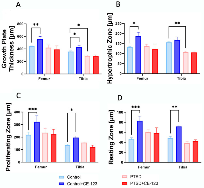
Background/Objectives: Post-traumatic stress disorder (PTSD) has been associated with negative effects on bone health, potentially leading to reduced bone mass, altered geometry, and impaired mechanical strength. However, the extent of these changes and possible pharmacological interventions remains unclear. This study aimed to assess the impact of PTSD on bone properties and evaluate the therapeutic potential of CE-123 in mitigating PTSD-induced skeletal deterioration. Additionally, we examined the effects of CE-123 on healthy bone to determine its broader influence on skeletal integrity and growth. Methods: We conducted an experiment using female Wistar rats divided into four groups: Control, PTSD, Control+CE-123, and PTSD+CE-123. PTSD was induced using a validated stress paradigm, and CE-123 was administered to evaluate its effects on bone properties. Morphometric, densitometric, and mechanical parameters of the tibia and femur were analyzed, along with growth plate measurements to assess potential effects on skeletal development. Results: PTSD led to significant reductions in bone mineral density, bone mass, and mechanical properties, particularly in cortical thickness and relative bone weight, suggesting increased bone fragility. CE-123 treatment in PTSD-exposed rats prevented some of these adverse effects but did not fully restore bone integrity. In healthy rats, CE-123 increased bone length and growth plate size, particularly in the proliferative and resting zones, indicating a stimulatory effect on bone growth. Conclusions: PTSD negatively affects bone structure and mechanical strength, while CE-123 shows a potential to mitigate these effects. However, its influence on healthy bones raises questions about its long-term impact on skeletal development. Further studies are needed to evaluate CE-123's clinical applicability and safety, particularly in younger populations.

Keywords: chronic stress; dopaminergic modulation; femur; growth plate; tibia.


Conflict of interest statement

The authors declare no conflicts of interest.

Figures

Figure 1
Figure 1
Comparison of femur and tibia morphological characteristics (body weight used as reference for other measurements) among four experimental groups: Control, PTSD, Control treated with CE-123, and PTSD treated with CE-123. Measured parameters include (A) body weight, (B) bone weight, (C) relative bone weight (RBW), (D) bone length. Asterisks (*) indicate significant differences between groups (* p < 0.05, ** p < 0.01, *** p < 0.001).
Figure 2
Figure 2
Comparison of femur and tibia bone properties among four experimental groups: Control, PTSD, Control treated with CE-123, and PTSD treated with CE-123. Measured parameters include (A) bone mineral density (BMD), (B) bone mineral content (BMC), (C) Seedor index. Asterisks (*) indicate significant differences between groups (* p < 0.05, ** p < 0.01).
Figure 3
Figure 3
Comparison of femur and tibia geometrical properties among four experimental groups: Control, PTSD, Control treated with CE-123, and PTSD treated with CE-123. Measured parameters include (A) transversal outer diameter (Hout), (B) transversal inner diameter (Hinn), (C) cranial–caudal outer diameter (Vout), (D) cranial–caudal inner diameter (Vinn), (E) mid-diaphysis cross-sectional area (CSA), (F) mean relative wall thickness (MRWT), (G) cortical index (CI), (H) cross-sectional moment of inertia (Ix). Asterisks (*) indicate significant differences between groups (* p < 0.05, ** p < 0.01, *** p < 0.001).
Figure 4
Figure 4
Comparison of femur and tibia mechanical properties among four experimental groups: Control, PTSD, Control treated with CE-123, and PTSD treated with CE-123. Measured parameters include (A) yield force (Fyield), (B) elastic work (Wyield), (C) stiffness, (D) breaking force (Fmax), (E) breaking work (Wmax). Asterisks (*) indicate significant differences between groups (* p < 0.05, ** p < 0.01).
Figure 5
Figure 5
Comparison of femur and tibia bone material properties among four experimental groups: Control, PTSD, Control treated with CE-123, and PTSD treated with CE-123. Measured parameters include (A) yield strain (Ɛyield), (B) yield stress (σyield), (C) breaking strain (Ɛmax), (D) breaking stress (σmax), (E) Young’s modulus. Asterisks (*) indicate significant differences between groups (* p < 0.05, *** p < 0.001).
Figure 6
Figure 6
A comparison of femur and tibia bone growth plate morphology among the four experimental groups: Control, PTSD, Control treated with CE-123, and PTSD treated with CE-123. The measurements of the growth plate include (A) overall growth plate thickness, (B) hypertrophic zone thickness, (C) proliferating zone thickness, and (D) resting zone thickness. Asterisks (*) indicate significant differences between the groups (* p < 0.05, ** p < 0.01, *** p < 0.001).
Figure 7
Figure 7
Representative microphotographs of the femoral growth plate (first column) and tibial growth plate (second column) in the Control group (first row), Control + CE-123 group (second row), PTSD group (third row), and PTSD + CE-123 group (fourth row). Letters indicate specific growth plate zones: r—resting zone; p—proliferative zone; h—hypertrophic zone. Scale bar = 100 µm.

Similar articles

References

    1. Messman T.L., LaPlena N., Wilensky S. Trauma-Related Disorders and Posttraumatic Stress Disorder. In: Friedman H.S., Markey C.H., editors. Encyclopedia of Mental Health. 3rd ed. Academic Press; Oxford, UK: 2023. pp. 501–510.
    1. Shalev A.Y. Posttraumatic Stress Disorder and Stress-Related Disorders. Psychiatr. Clin. N. Am. 2009;32:687–704. doi: 10.1016/j.psc.2009.06.001. - DOI - PMC - PubMed
    1. Chen X. The Causes and Effects of Post-Traumatic Stress Disorder. SHS Web Conf. 2023;157:04029. doi: 10.1051/shsconf/202315704029. - DOI
    1. Palmer S.J. The Complex Neurology That Links Physical Health to Post-Traumatic Stress Disorder. Br. J. Neurosci. Nuesing. 2023;19:37–39. doi: 10.12968/bjnn.2023.19.1.37. - DOI
    1. Da Costa Silva L., Laisney M., Charretier L., Eustache F., Quinette P. Memory alterations in post-traumatic stress disorder. Biol. Aujourdhui. 2023;217:55–64. doi: 10.1051/jbio/2023018. - DOI - PubMed

LinkOut - more resources