Review
. 2025 Mar 31;13(7):775.
doi: 10.3390/healthcare13070775.

The Evaluation, Diagnosis, and Management of Ovarian Cysts, Masses, and Their Complications in Fetuses, Infants, Children, and Adolescents

Affiliations
Review

The Evaluation, Diagnosis, and Management of Ovarian Cysts, Masses, and Their Complications in Fetuses, Infants, Children, and Adolescents

Marko Bašković et al. Healthcare (Basel). .

Abstract

The majority of abdominal masses in female children derive from the ovaries. Ovarian masses in pediatric populations can vary from simple functional cysts to malignant neoplasms. Their incidence, clinical presentation, and histological distribution vary across age groups. In the assessment of ovarian masses in children, the primary aim is to determine the probability of malignancy, as the treatment approaches for benign and malignant lesions are significantly distinct. The primary imaging tool for evaluating ovarian cysts and masses is ultrasound, which can assess the size, location, and characteristics of masses. Magnetic resonance imaging (MRI) or computed tomography (CT) may be used for further evaluation if ultrasound findings are inconclusive or if malignancy is suspected, especially in older adolescents. Serum markers may be considered in older adolescents to help assess the risk of malignancy, though it is less useful in younger populations due to normal developmental variations. Many functional ovarian cysts, especially those detected in fetuses or infants, often resolve spontaneously without intervention. Surgical intervention is indicated in cases of large cysts that cause symptoms, or if there are concerns for malignancy. Common procedures include primarily ovarian sparing laparoscopy or laparotomy. Complications like torsion, rupture, or hemorrhage may require urgent surgical intervention. Treatment should be performed in specialized centers to avoid unnecessary oophorectomies and ensure the best possible outcome for the patient. This comprehensive review aims to provide an overview of the evaluation, diagnosis, and treatment of ovarian masses in the pediatric population. Emphasis is placed on the particularities of the lesions and their management in relation to age subgroups.

Keywords: adolescent; children; cystectomy; fetus; gynecology; infant; oophorectomy; ovarian cyst; ovarian mass; ovary; pediatric surgery.


Conflict of interest statement

The authors declare no conflicts of interest.

Figures

Figure 1
Figure 1
A 7-day-old female neonate presented with a large abdominal mass. (A) Laparoscopic examination revealed a necrotic, twisted, huge neonatal ovarian cyst measuring 6 × 5 × 4 cm; (B) Twisting of the ovarian pedicle by 720 degrees; (C) After decompression of the cyst, a laparoscopic salpingo-oophorectomy was performed due to the obvious necrosis of the ovarian tissue. Source: Archive of the Department of Pediatric Surgery, University Hospital of Split.
Figure 2
Figure 2
A 13-year-old adolescent presented to the emergency room because of recurrent crampy abdominal pain that lasted a short time and then subsided. She had had the symptoms for about a week. An abdominal ultrasound was performed, which showed a mass in the pelvis. (A) MSCT of the abdomen revealed a 9.1 × 8.8 cm solid cystic mass in the right ovary that was not perfused; (B) Intraoperatively, a torquing, partially necrotic tumor of the ovary was noted; (C) Pathohistological findings were consistent with a mature teratoma of the right ovary. Source: Archive of the Department of Pediatric Surgery, University Hospital of Split.
Figure 3
Figure 3
A 14-year-old girl presented with intermittent abdominal pain, loss of appetite, and a palpable abdominal mass. (A) Multislice computed tomography revealed a giant right abdominal ovarian cyst measuring 16.2 × 14.6 cm; (B) The cystic mass filled the entire lower abdomen and was pressing on the surrounding structures; (C) Laparoscopic examination revealed a giant ovarian cyst; (D) The ovarian cortex was opened, 2 L of clear contents were aspirated; (E) A laparoscopic cystectomy with ovarian sparing was performed. A pathohistological examination revealed a simple ovarian cyst. Source: Archive of the Department of Pediatric Surgery, University Hospital of Split.
Figure 4
Figure 4
An 11-year-old girl noticed that her stomach was growing along with pain, nausea, and a palpable mass. Ultrasound and MRI revealed bilateral ovarian tumor inhomogeneous solid cystic masses similar to dermoid, left 13 cm, right 5 cm in diameter. Tumor markers were not increased, and due to the size of the tumor, a laparotomy approach was decided. Intraoperative findings: bilateral asymmetrical ovarian masses and bilateral cystectomy were performed. A pathohistological examination revealed a mature teratoma. Source: Archive of the Department of Obstetrics and Gynecology, Clinical Hospital Merkur.
Figure 5
Figure 5
A 17-year-old female patient presented to a pediatric endocrinologist for primary amenorrhea. After diagnostic evaluation, the endocrinologist diagnosed a female phenotype and a male karyotype (46, XY). The MR scan of the abdomen showed a tumor in both gonads. The left gonad looked like a testis and the right one like an ovary. A diagnosis of a disorder of sexual development (DSD) was made. A bilateral laparoscopic adnexectomy was performed. The pathohistological examination revealed a bilateral gonadoblastoma with components of a dysgerminoma. No gonadal tissue was present in either of the removed specimens. TNM: T1bNXMX; Figo classification: 1b; (A) Intraoperative findings; (B) Macroscopic specimen after bilateral adnexectomy. Source: Archive of the Department of Pediatric Surgery, University Hospital of Split.
Figure 6
Figure 6
A 14-year-old girl presented with abdominal distension that had been present for several months. Apart from occasional constipation, she had no other symptoms. (A) MRI of the abdomen shows an intraperitoneal mass inseparable from the right ovary, compressing the pancreas and abdominal aorta; (B) Surgical exploration of the abdomen—mature cystic ovarian teratoma with foci of adenocarcinomas measuring 28 × 22 × 14 cm. Source: Archive of the Department of Pediatric Surgery, Children’s Hospital Zagreb.
Figure 7
Figure 7
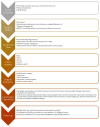
Flowchart for management of pediatric ovarian masses.
Figure 8
Figure 8
A 14-year-old girl presented with acute abdominal pain and vomiting. Abdominal ultrasonography revealed a left ovarian cystic-solid mass 11 × 8 cm with destruction and free fluid in the abdomen. Because of acute abdomen, exploratory laparotomy and left-side adnexectomy were performed. A ruptured torquated ovarian tumor was found. A pathohistological examination revealed a ruptured ovarian cystic mature teratoma. Source: Archive of the Department of Obstetrics and Gynecology, Clinical Hospital Merkur.
Figure 9
Figure 9
A 15-year-old girl presented with abdominal pain, fever and vomiting. The symptoms started five days prior to surgery. Abdominal ultrasonography revealed a huge cystic mass in the pelvis measuring 13 × 10 × 8 cm adjacent to the left ovary, which was edematous and without visible blood flow. (A) A laparoscopic examination revealed a huge paraovarian cyst and torsion of the left adnexa; (B) A laparoscopic salpingo-oophorectomy was performed due to the obvious gangrene of the left ovary and fallopian tube. A pathohistological examination revealed a necrotic paraovarian cyst and gangrene of the left ovary. Source: Archive of the Department of Pediatric Surgery, University Hospital of Split.

Similar articles

Cited by

References

    1. Ciro E., Vincenzo C., Mariapina C., Fulvia D.C., Vincenzo B., Giorgia E., Roberto C., Lepore B., Castagnetti M., Califano G., et al. Review of a 25-Year Experience in the Management of Ovarian Masses in Neonates, Children and Adolescents: From Laparoscopy to Robotics and Indocyanine Green Fluorescence Technology. Children. 2022;9:1219. doi: 10.3390/children9081219. - DOI - PMC - PubMed
    1. Birbas E., Kanavos T., Gkrozou F., Skentou C., Daniilidis A., Vatopoulou A. Ovarian Masses in Children and Adolescents: A Review of the Literature with Emphasis on the Diagnostic Approach. Children. 2023;10:1114. doi: 10.3390/children10071114. - DOI - PMC - PubMed
    1. Lam C.Z., Chavhan G.B. Magnetic resonance imaging of pediatric adnexal masses and mimics. Pediatr. Radiol. 2018;48:1291–1306. doi: 10.1007/s00247-018-4073-4. - DOI - PubMed
    1. Heo S.H., Kim J.W., Shin S.S., Jeong S.I., Lim H.S., Choi Y.D., Lee K.H., Kang W.D., Jeong Y.Y., Kang H.K. Review of ovarian tumors in children and adolescents: Radiologic-pathologic correlation. Radiographics. 2014;34:2039–2055. doi: 10.1148/rg.347130144. - DOI - PubMed
    1. Garel L., Dubois J., Grignon A., Filiatrault D., Van Vliet G. US of the pediatric female pelvis: A clinical perspective. Radiographics. 2001;21:1393–1407. doi: 10.1148/radiographics.21.6.g01nv041393. - DOI - PubMed

LinkOut - more resources