

Optical Coherence Tomography in Retinal Detachment: Prognostic Biomarkers, Surgical Planning, and Postoperative Monitoring
- PMID: 40218221
- PMCID: PMC11988441
- DOI: 10.3390/diagnostics15070871
Optical Coherence Tomography in Retinal Detachment: Prognostic Biomarkers, Surgical Planning, and Postoperative Monitoring
Abstract
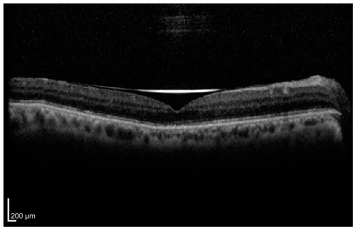
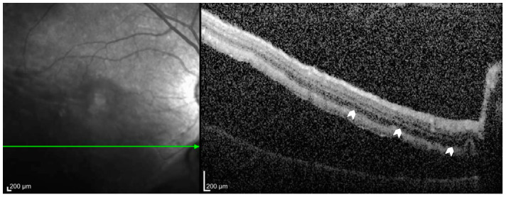
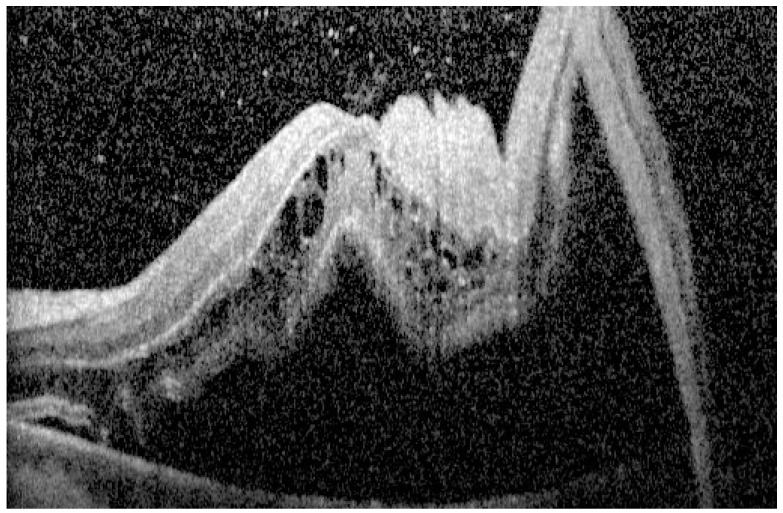
Retinal detachment (RD) is a vision-threatening ocular emergency that necessitates rapid diagnosis and intervention. This review examines the evolving role of optical coherence tomography (OCT) in RD by synthesizing the literature on preoperative biomarkers and advanced image modalities that inform diagnosis, prognosis, and surgical planning. We evaluated studies employing spectral-domain OCT, swept-source OCT, OCT angiography, adaptive optics OCT, and en face OCT to assess photoreceptor integrity, retinal detachment height, intraretinal cystic cavities, outer retinal corrugations and undulation, and macular involvement. The incorporation of OCT assessment into clinical practice may facilitate more precise surgical timing, technique selection, and postoperative monitoring. Further research is needed to standardize imaging protocols and validate specific prognostic biomarkers for optimal surgical outcomes. Key aspects include uniform imaging protocols, validating OCT-derived biomarkers such as ellipsoid zone integrity, and correlating OCT metrics with functional outcomes.
Keywords: OCT angiography; adaptive optics OCT; en face OCT; optical coherence tomography; photoreceptor integrity; prognostic biomarkers; retinal detachment; surgical planning; swept-source OCT.
Conflict of interest statement
The authors declare no conflicts of interest.
Figures
Similar articles
-
BACILLARY LAYER DETACHMENT OVERLYING REDUCED CHORIOCAPILLARIS FLOW IN ACUTE IDIOPATHIC MACULOPATHY.Retin Cases Brief Rep. 2022 Jan 1;16(1):59-66. doi: 10.1097/ICB.0000000000000943. Retin Cases Brief Rep. 2022. PMID: 31764886
-
[Macula analysis by spectral domain OCT in rhegmatogenous retinal detachment surgery].J Fr Ophtalmol. 2015 Mar;38(3):181-92. doi: 10.1016/j.jfo.2014.09.015. Epub 2015 Feb 4. J Fr Ophtalmol. 2015. PMID: 25662918 French.
-
Structural recovery of the detached macula after retinal detachment repair as assessed by optical coherence tomography.Korean J Ophthalmol. 2013 Jun;27(3):178-85. doi: 10.3341/kjo.2013.27.3.178. Epub 2013 May 7. Korean J Ophthalmol. 2013. PMID: 23730110 Free PMC article.
-
Optical Coherence Tomography Findings in Rhegmatogenous Retinal Detachment: A Systematic Review.J Clin Med. 2022 Sep 30;11(19):5819. doi: 10.3390/jcm11195819. J Clin Med. 2022. PMID: 36233684 Free PMC article. Review.
-
[Optical coherence tomography biomarkers for neovascular age-related macular degeneration : Relevance for the diagnosis, treatment and prognosis].Ophthalmologie. 2025 Feb;122(2):144-156. doi: 10.1007/s00347-024-02181-8. Epub 2025 Jan 27. Ophthalmologie. 2025. PMID: 39870835 Review. German.
References
Publication types
LinkOut - more resources
Full Text Sources
Research Materials
Miscellaneous
