

Preliminary Exploration of MAGE-B1, -B4, -B5, and -B10 mRNA Expression in Canine Mammary Tumors in Dogs
- PMID: 40218304
- PMCID: PMC11987965
- DOI: 10.3390/ani15070910
Preliminary Exploration of MAGE-B1, -B4, -B5, and -B10 mRNA Expression in Canine Mammary Tumors in Dogs
Abstract
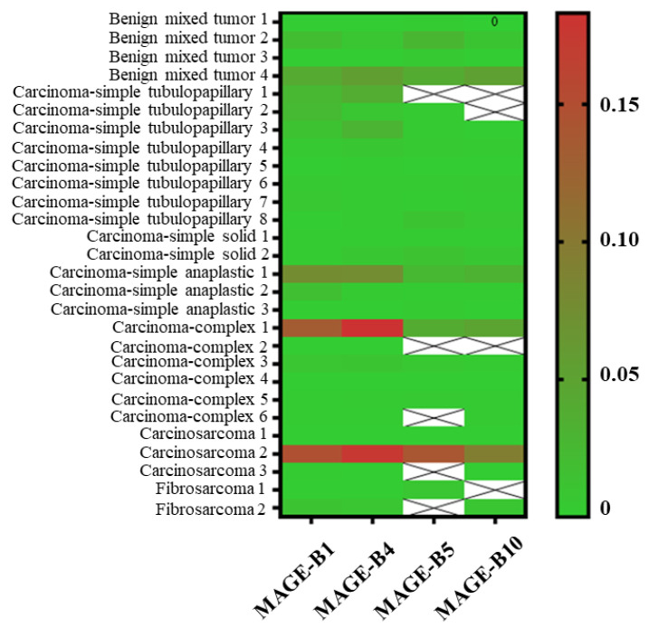
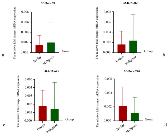
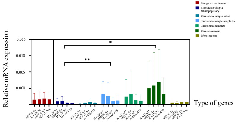
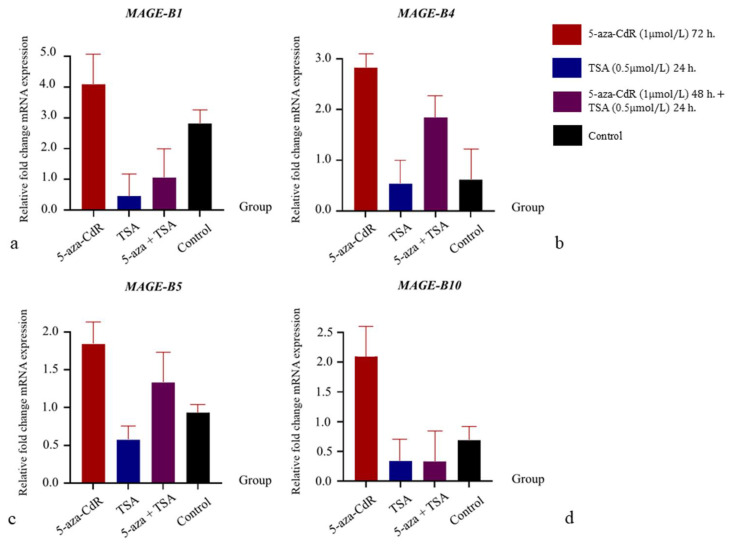
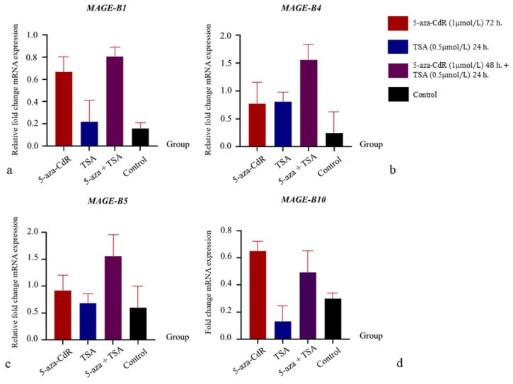
The melanoma-associated antigen gene (MAGE) is a key target in cancer immunotherapy. Given the potential of MAGE-B genes in veterinary immunotherapy for canine mammary tumors (CMTs), this study investigated the mRNA expression of MAGE-B1, -B4, -B5, and -B10 in CMT tissues and cells from dogs. Quantitative real-time PCR was used to analyze 28 CMT tissue samples, including 4 benign and 24 malignant tumors (13 simple carcinomas, 6 complex carcinomas, 3 carcinosarcomas, and 2 fibrosarcomas). Benign mixed tumor and complex carcinoma-type CMT cells were cultured and treated with a DNA methylase inhibitor (5-aza-2'-deoxycytidine; 5-aza-CdR) and a histone deacetylase inhibitor (Trichostatin A; TSA) under the following four conditions: (1) 5-aza-CdR for 72 h; (2) TSA for 24 h; (3) 5-aza-CdR for 48 h followed by TSA for 24 h; and (4) control. MAGE-B1 and -B4 showed the highest expression in the CMT samples (100% and 89.29%, respectively), followed by MAGE-B10 (82.14%). Carcinosarcomas and simple anaplastic carcinomas had significantly higher MAGE-B expression levels than simple tubulopapillary carcinomas (p < 0.05). 5-aza-CdR treatment increased MAGE-B expression, whereas TSA had a mild effect. Further research involving larger cohorts is needed to confirm these findings.
Keywords: canine mammary tumors; mRNA expression; melanoma-associated antigen-B; quantitative real-time PCR.
Conflict of interest statement
The authors declare no conflicts of interest. The funders had no role in the design of the study; in the collection, analyses, or interpretation of data; in the writing of the manuscript; or in the decision to publish the results.
Figures
Similar articles
-
[Expression of melanoma-associated antigen-C2 in breast cancers and mechanism].Zhonghua Zhong Liu Za Zhi. 2021 Aug 23;43(8):821-826. doi: 10.3760/cma.j.cn112152-20200116-00043. Zhonghua Zhong Liu Za Zhi. 2021. PMID: 34407585 Chinese.
-
A Preliminary Study of the Cross-Reactivity of Canine MAGE-A with Hominid Monoclonal Antibody 6C1 in Canine Mammary Gland Tumors: An Attractive Target for Cancer Diagnostic, Prognostic and Immunotherapeutic Development in Dogs.Vet Sci. 2020 Aug 10;7(3):109. doi: 10.3390/vetsci7030109. Vet Sci. 2020. PMID: 32784970 Free PMC article.
-
Expressions of MAGE-A9 and MAGE-A11 in breast cancer and their expression mechanism.Arch Med Res. 2014 Jan;45(1):44-51. doi: 10.1016/j.arcmed.2013.10.005. Epub 2013 Dec 5. Arch Med Res. 2014. PMID: 24316396
-
The expression of MAGE-C1 and MAGE-C2 in breast cancer and their clinical significance.Am J Surg. 2016 Jan;211(1):142-51. doi: 10.1016/j.amjsurg.2015.05.028. Epub 2015 Aug 3. Am J Surg. 2016. PMID: 26321295
-
Know Thy Model: Charting Molecular Homology in Stromal Reprogramming Between Canine and Human Mammary Tumors.Front Cell Dev Biol. 2019 Dec 17;7:348. doi: 10.3389/fcell.2019.00348. eCollection 2019. Front Cell Dev Biol. 2019. PMID: 31921858 Free PMC article. Review.
References
-
- Misdorp W. Tumors of the mammary gland. In: Meuten D.J., editor. Tumors in Domestic Animals. 1st ed. Blackwell Press; Oxford, UK: 2002. pp. 575–606.
-
- Srisawat W., Pringproa K., Prachasilchai W., Thongtharb A., Sthitmatee N. Epidemiology and classification for canine and feline mammary gland tumors: A histopathological survey of 437 mammary gland tumor biopsies performed in a secondary care hospital in Chiang Mai, Thailand from 2012 to 2019. PeerJ. 2024;12:e17077. - PMC - PubMed
-
- Lana S.E., Rutteman G.R., Withrow S.J. Tumors of the mammary gland. In: Withrow S.J., Vail D.M., editors. Small Animal Clinical Oncology. 1st ed. Elsevier Press; St. Louis, MO, USA: 2007. pp. 619–636.
-
- Saba C.F., Rogers K.S., Newman S.J., Mauldin G.E., Vail D.M. Mammary gland tumors in male dogs. J. Vet. Intern. Med. 2007;21:1056–1059. - PubMed
Grants and funding
LinkOut - more resources
Full Text Sources
Research Materials
