. 2025 Mar 23;15(7):923.
doi: 10.3390/ani15070923.

Repurposing Vancomycin as a Potential Antiviral Agent Against PEDV via nsp13 Helicase Inhibition

Affiliations

Repurposing Vancomycin as a Potential Antiviral Agent Against PEDV via nsp13 Helicase Inhibition

Qiao Chen et al. Animals (Basel). .

Abstract

Porcine epidemic diarrhea virus (PEDV) causes a highly contagious intestinal disease with severe economic impacts on the global swine industry. The non-structural protein 13 (nsp13), a viral helicase, is essential for viral replication, making it a promising target for antiviral drug development. In this study, through virtual screening and molecular dynamics simulations, Vancomycin, a small-molecule drug also clinically used as an antibacterial agent, was identified to exhibit a stable binding affinity for PEDV nsp13. The NTPase and ATP-dependent RNA helicase activities of PEDV nsp13 were confirmed in vitro, and the optimal biochemical reaction conditions for its dsRNA unwinding activity were established. Further experiments demonstrated that Vancomycin effectively inhibited the dual enzymatic activities of PEDV nsp13 and reduced PEDV infections in vitro. This research highlights Vancomycin as a novel inhibitor of PEDV nsp13, providing valuable mechanistic insights and serving as a model for antiviral drug discovery. While this study suggests its potential for repurposing as a therapeutic agent against PEDV, further investigations are required to evaluate its feasibility in vivo, particularly in terms of safety, efficacy, and practical applicability.

Keywords: antiviral drug; helicase inhibitor; nsp13; porcine epidemic diarrhea virus; vancomycin; virtual screening.


Conflict of interest statement

The authors declare no conflicts of interest.

Figures

Figure 1
Figure 1
Virtual screening analysis of drugs targeting PEDV nsp13. (A) Binding mode of Vancomycin within the active pocket of the PEDV nsp13 protein. (1) Homology model of nsp13 and Vancomycin complex, with nsp13 represented as a gray cartoon, Vancomycin as yellow sticks, and key interacting residues as green sticks. (2) A 3D visualization of the Vancomycin–nsp13 binding mode. (3) A 2D interaction map highlighting key residues and interactions, including hydrogen bonds and hydrophobic forces. (BD) Molecular dynamics (MD) simulation results. (B) Root mean square deviation (RMSD) of the backbone of nsp13 over 100 ns. (C) Radius of gyration (Rg) of the Vancomycin–nsp13 complex, indicating structural compactness. (D) Number of hydrogen bonds formed between Vancomycin and nsp13 during the simulation.
Figure 2
Figure 2
PEDV nsp13 exhibits NTPase activities. (A) The purified nsp13 protein was analyzed by 10% SDS-PAGE and stained with Coomassie brilliant blue (left) or subjected to Western blot with an anti-His antibody (right). (B) 1 nM PEDV nsp13 was incubated with various NTPs (2.5 mM each), and NTPase activity was measured as nanomoles of released inorganic phosphate (Pi) using the Malachite Green Phosphate Detection Kit. The reaction without any NTP was used as a negative control (none). (C) ATPase activity of PEDV nsp13 was assessed by incubating 2.5 mM ATP with increasing concentrations of nsp13. (D) ATPase activity was tested with 2.5 mM ATP and 2 mM of various divalent metal ions. The reaction without metal ions was used as a negative control (none). (E) ATP hydrolysis was measured with increasing concentrations of Mg2+. Error bars represent the standard deviation (SD) from three separate experiments.
Figure 3
Figure 3
PEDV nsp13 exhibits RNA helicase activity. (A) Schematic of the RNA duplex substrate (RNA*/RNA). Asterisks indicate the Cy5-labeled strands. (B) The RNA duplex substrate (2 nM) was incubated with 2 μM nsp13, and unwinding was analyzed via gel electrophoresis and imaged on a Typhoon imager. Negative controls included non-boiled and ATP-free reaction mixtures, while a boiled reaction mixture (95 °C) served as the positive control. (C) The RNA duplex unwinding assay was conducted with increasing concentrations of PEDV nsp13. (D) Time-course analysis of RNA unwinding, with 2 μM nsp13 and 2 nM RNA substrate monitored over varying reaction times.
Figure 4
Figure 4
Biochemical analysis of the optimal RNA duplex unwinding activity of PEDV Nsp13. Asterisks indicate the Cy5-labeled strands. (A) PEDV nsp13 (2 μM) was reacted with the RNA duplex substrate (2 nM) under different biochemical conditions, including the indicated NTPs (2 mM), (B) increasing concentrations of ATP, (C) each indicated divalent metal ion (2 mM), (D) increasing concentrations of Mg2+, (E) varying reaction temperatures for 30 min, and (F) different reaction pH values. The unwinding activity was assessed as described above.
Figure 5
Figure 5
Vancomycin directly inhibited the ATPase activity and RNA duplex unwinding activity of PEDV nsp13. Asterisks indicate the Cy5-labeled strands. (A) PEDV nsp13 (2 μM) was incubated with 2.5 mM ATP at increasing concentrations of Vancomycin (0, 0.5, 1, 2, 5, and 10 μM), and ATPase activity was measured as described above. (B) PEDV nsp13 (2 μM) was reacted with an RNA duplex substrate (2 nM) under increasing concentrations of Vancomycin (0, 0.5, 1, 2, 5, and 10 μM), and the RNA duplex unwinding activity was assessed as described above. (C) The unwinding activity was analyzed using ImageJ (version 1.47v, National Institutes of Health, Bethesda, MD, USA) and plotted as the percentage of Cy5-labeled RNA released from the total RNA duplex (Y-axis) at the indicated concentrations of Vancomycin (X-axis). Error bars represent significant differences (*** p < 0.001; ns = not significant).
Figure 6
Figure 6
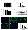
Vancomycin exhibits antiviral activity against PEDV CV777 strains in Vero cells. (A) Cell viability of Vero cells treated with the indicated concentrations of Vancomycin. (BE) Vero cells were treated with the specified concentrations of Vancomycin or DMSO as a negative control and then infected with PEDV at an MOI of 0.01, and samples were collected after 12 h. (B) Relative levels of PEDV N mRNA were quantified by qRT-PCR, normalized to GAPDH, and expressed relative to levels in DMSO-treated cells. (C) PEDV (CV777 strain) viral titers were determined in Vancomycin-treated and DMSO-treated Vero cells using the TCID50 assay. (D) PEDV N-protein levels were evaluated by Western blot, with GAPDH as the internal control. (E) Levels of PEDV infection in Vero cells were assessed using immunofluorescence, with PEDV N-protein shown in green. Scale bar = 500 μm. Error bars represent SD values from three independent experiments. Asterisks in the figures indicate significant differences (** p < 0.01; *** p < 0.001; ns = not significant).
Figure 7
Figure 7
Antiviral effect of Vancomycin on PEDV. (A) Cell viability was measured under a series of Vancomycin concentrations using the CCK-8 assay, and the 50% Cytotoxic Concentration (CC50) values of Vancomycin were calculated based on the dose-response curve using GraphPad Prism 5. (B) PEDV CV777 was added at a multiplicity of infection (MOI) of 0.01 with a series of Vancomycin concentrations. Cell viability was measured, and the PEDV inhibition ratio was calculated at 24 h of Vancomycin treatment. The 50% Effective Concentration (EC50) values of Vancomycin were calculated using GraphPad Prism10 to assess inhibition ratios at different inhibitor concentrations. The error bars show the SD of the results from three replicates.

Similar articles

References

    1. Pensaert M.B., de Bouck P. A new coronavirus-like particle associated with diarrhea in swine. Arch. Virol. 1978;58:243–247. doi: 10.1007/BF01317606. - DOI - PMC - PubMed
    1. Zhang H., Zou C., Peng O., Ashraf U., Xu Q., Gong L., Fan B., Zhang Y., Xu Z., Xue C., et al. Global Dynamics of Porcine Enteric Coronavirus PEDV Epidemiology, Evolution, and Transmission. Mol. Biol. Evol. 2023;40:msad052. doi: 10.1093/molbev/msad052. - DOI - PMC - PubMed
    1. Ferrara G., Nocera F.P., Longobardi C., Ciarcia R., Fioretti A., Damiano S., Iovane G., Pagnini U., Montagnaro S. Retrospective Serosurvey of Three Porcine Coronaviruses among the Wild Boar (Sus scrofa) Population in the Campania Region of Italy. J. Wildl. Dis. 2022;58:887–891. doi: 10.7589/JWD-D-21-00196. - DOI - PubMed
    1. Ferrara G., D’Anza E., Rossi A., Improda E., Iovane V., Pagnini U., Iovane G., Montagnaro S. A Serological Investigation of Porcine Reproductive and Respiratory Syndrome and Three Coronaviruses in the Campania Region, Southern Italy. Viruses. 2023;15:300. doi: 10.3390/v15020300. - DOI - PMC - PubMed
    1. Gerdts V., Zakhartchouk A. Vaccines for porcine epidemic diarrhea virus and other swine coronaviruses. Vet. Microbiol. 2017;206:45–51. doi: 10.1016/j.vetmic.2016.11.029. - DOI - PMC - PubMed

LinkOut - more resources