. 2025 Mar 30;17(7):1217.
doi: 10.3390/nu17071217.

Dual Action of Pueraria montana var. lobata Extract on Myogenesis and Muscle Atrophy

Affiliations

Dual Action of Pueraria montana var. lobata Extract on Myogenesis and Muscle Atrophy

So Young Eun et al. Nutrients. .

Abstract

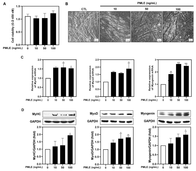
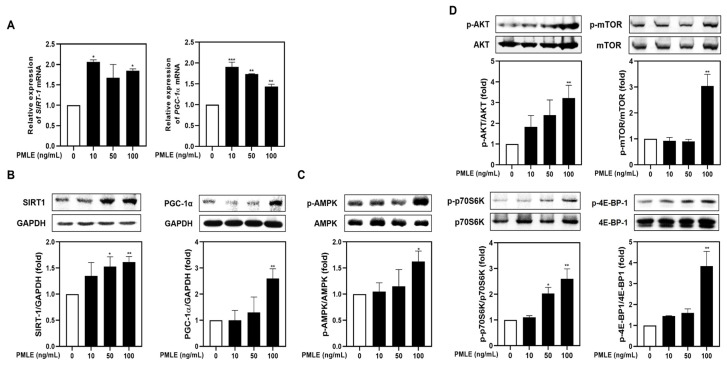
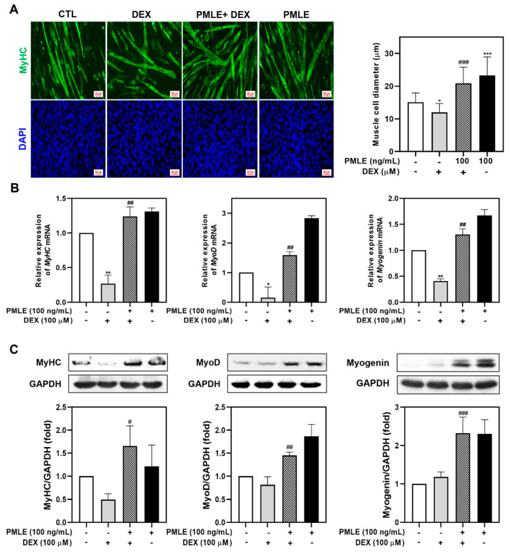
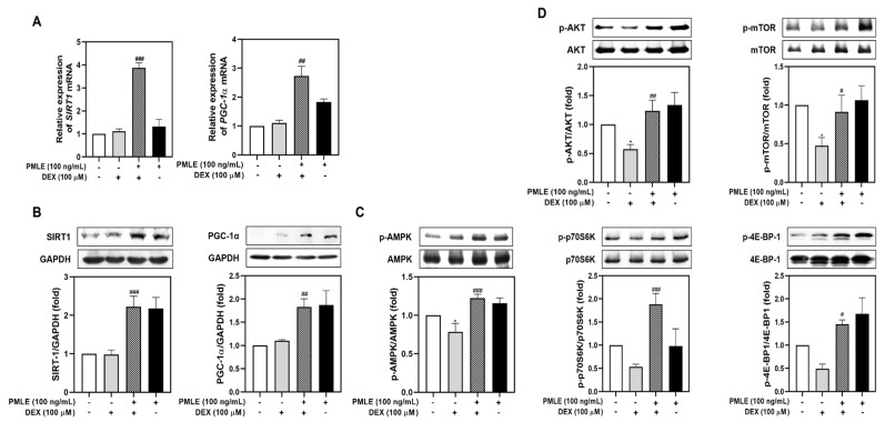
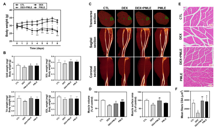
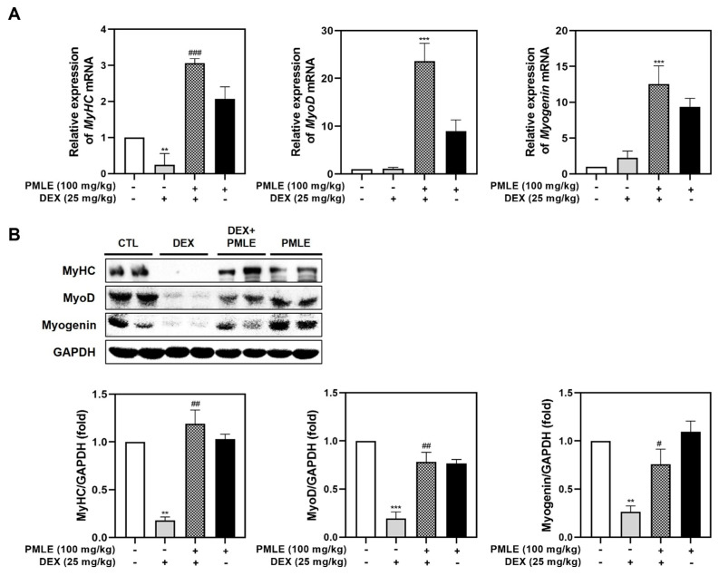
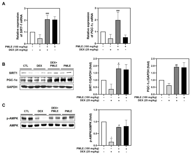
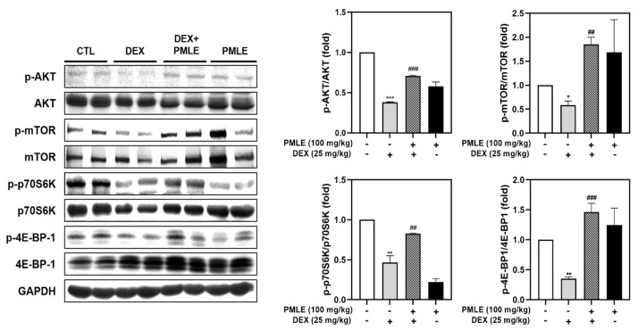
Background/Objectives: Muscle atrophy, defined by diminished muscle mass and function, is a notable concern associated with aging, disease, and glucocorticoid treatment. Pueraria montana var. lobata extract (PMLE) demonstrates multiple bioactive properties, such as antioxidant, anti-inflammatory, and metabolic regulatory activities; however, its role in muscle atrophy has not been extensively investigated to date. This study examined how PMLE influences both muscle cell differentiation and dexamethasone (DEX)-induced muscle degeneration by focusing on the underlying molecular mechanisms. Methods: This study examined the effects of PMLE on myogenic differentiation and DEX-induced muscle atrophy. C2C12 myoblasts were treated with PMLE (10-100 ng/mL) and assessed for changes in the expression of myogenesis-related genes and activation of Akt/mTOR and AMPK/SIRT1/PGC-1α signaling cascades. In vivo, a DEX-induced muscle atrophy model was used to assess muscle mass, fiber morphology, and molecular changes. Results: PMLE PMLE promoted muscle cell development by increasing the expression of MyHC, MyoD, and myogenin while activating protein synthesis and mitochondrial biogenesis pathways. PMLE counteracted DEX-induced myotube atrophy, restoring myotube diameter and promoting cellular fusion in vitro. In vivo, PMLE mitigated muscle degradation in fast-twitch muscle groups and reversed DEX-induced suppression of key anabolic and mitochondrial pathways. Conclusions: These findings suggest that PMLE promotes myogenic differentiation and protects against muscle atrophy by regulating critical molecular pathways, indicating its promise as a treatment candidate for conditions involving muscle wasting. Further studies are required to assess its clinical application and long-term safety efficacy.

Keywords: Pueraria montana var. lobata extract; dexamethasone; mitochondrial biogenesis; muscle atrophy; protein synthesis.


Conflict of interest statement

The authors declare no conflicts of interest.

Figures

Figure 1
Figure 1
Effects of Pueraria montana var. lobata extract (PMLE) on myogenic differentiation in C2C12 myoblasts. (A) XTT assay showing no significant effect of PMLE (10, 50, 100 ng/mL) on C2C12 cell viability. Data are mean ± SD. (B) Phase-contrast images of myotube formation after PMLE treatment. Scale bar = 50 µm. (C) qRT-PCR analysis of MyHC, MyoD, and myogenin mRNA levels after PMLE treatment. Data are normalized to GAPDH and shown as fold change vs. control. (D) Western blot analysis of MyHC, MyoD, and myogenin protein levels. GAPDH was used as a loading control. Band intensities are shown as fold change vs. control. Statistical significance: * p < 0.05, ** p < 0.01, *** p < 0.001 vs. control.
Figure 2
Figure 2
PMLE enhances mitochondrial biogenesis and protein synthesis signaling pathways in C2C12 myoblasts. (A) qRT-PCR analysis of SIRT1 and PGC-1α mRNA levels after PMLE (10, 50, 100 ng/mL) treatment. Data are normalized to GAPDH and shown as fold change vs. control. (B) Western blot analysis of SIRT1 and PGC-1α protein levels. GAPDH was used as a loading control. (C) Western blot of phosphorylated AMPK (p-AMPK); total AMPK was used as a control. (D) Western blot of p-AKT, p-mTOR, p-p70S6K, and p-4E-BP1; corresponding total proteins were used as loading controls. Band intensities are shown as fold change vs. control. Statistical significance: * p < 0.05, ** p < 0.01, *** p < 0.001 vs. control. PMLE, Pueraria montana var. lobata extract.
Figure 3
Figure 3
PMLE alleviates dexamethasone (DEX)-induced muscle atrophy and promotes myogenic differentiation in C2C12 myotubes. (A) Immunofluorescence staining of MyHC (green) and nuclei (DAPI, blue) in C2C12 myotubes treated with DEX (100 μM) and/or PMLE (100 ng/mL). The right panel shows the quantification of myotube diameter. (B) qRT-PCR analysis of MyHC, MyoD, and myogenin mRNA levels. Data are normalized to GAPDH and shown as fold change vs. control. (C) Western blot analysis of MyHC, MyoD, and myogenin protein levels. GAPDH was used as a loading control. Band intensities are shown as fold change vs. control. Statistical significance: * p < 0.05, ** p < 0.01, *** p < 0.001 vs. control group; # p < 0.05, ## p < 0.01, ### p < 0.001 vs. DEX-treated group. PMLE, Pueraria montana var. lobata extract.
Figure 4
Figure 4
PMLE restores mitochondrial biogenesis and protein synthesis signaling pathways in DEX-induced muscle atrophy. (A) qRT-PCR analysis of SIRT1 and PGC-1α mRNA levels in C2C12 myotubes treated with DEX (100 μM) and/or PMLE (100 ng/mL). Data are normalized to GAPDH and shown as fold change vs. control. (B) Western blot of SIRT1 and PGC-1α protein levels. GAPDH was used as a loading control. (C) Western blot of phosphorylated AMPK (p-AMPK); total AMPK was used as a control. (D) Western blot of p-AKT, p-mTOR, p-p70S6K, and p-4E-BP1; total proteins were used as loading controls. Band intensities are presented as fold change vs. control. Statistical significance: * p < 0.05 vs. control group; # p < 0.05, ## p < 0.01, ### p < 0.001 vs. DEX-treated group. PMLE, Pueraria montana var. lobata extract.
Figure 5
Figure 5
PMLE prevents DEX-induced muscle atrophy in vivo. (A) Body weight changes in mice treated with DEX (20 mg/kg) and/or PMLE (50 mg/kg) for 8 days. (B) Relative weights of GAS, SOL, TA, and EDL muscles normalized to body weight. (C) Micro-CT images (transverse, sagittal, coronal) of lower limb muscles. Green outlines indicate muscle boundaries. (D) Quantification of muscle volume and surface area (% of control). (E) H&E staining of GAS muscle cross-sections. Scale bar = 50 µm. (F) Quantitative analysis of myofiber CSA from histological images showing PMLE-mediated recovery of fiber size reduced by DEX treatment. Statistical significance: * p < 0.05, ** p < 0.01, *** p < 0.001 vs. control group; # p < 0.05, ## p < 0.01, ### p < 0.001 vs. DEX-treated group. PMLE, Pueraria montana var. lobata extract; CSA, cross-sectional area.
Figure 6
Figure 6
PMLE restores myogenic marker expression in DEX-induced muscle atrophy in vivo. (A) qRT-PCR analysis of MyHC, MyoD, and myogenin mRNA levels in gastrocnemius muscle from mice treated with DEX (25 mg/kg) and/or PMLE (100 mg/kg). Data are normalized to GAPDH and shown as fold change vs. control. (B) Western blot analysis of MyHC, MyoD, and myogenin protein levels in gastrocnemius muscle (CTL, DEX, DEX + PMLE, PMLE). GAPDH was used as a loading control. Band intensities are shown as fold change vs. control. Statistical significance: ** p < 0.01, *** p < 0.001 vs. control group; # p < 0.05, ## p < 0.01, ### p < 0.001 vs. DEX-treated group. PMLE, Pueraria montana var. lobata extract; DEX, dexamethasone; CTL, control.
Figure 7
Figure 7
PMLE restores mitochondrial biogenesis and energy metabolism-related signaling pathways in DEX-induced muscle atrophy in vivo. (A) qRT-PCR analysis of SIRT1 and PGC-1α mRNA levels in gastrocnemius muscle from mice treated with DEX (25 mg/kg) and/or PMLE (100 mg/kg). Data are normalized to GAPDH and shown as fold change vs. control. (B) Western blot analysis of SIRT1 and PGC-1α protein levels in each treatment group (CTL, DEX, DEX + PMLE, PMLE). GAPDH was used as a loading control. (C) Western blot of phosphorylated AMPK (p-AMPK); total AMPK used as a loading control. Band intensities are shown as fold change vs. control. Statistical significance: * p < 0.05 vs. control group; # p < 0.05, ## p < 0.01, ### p < 0.001 vs. DEX-treated group. PMLE, Pueraria montana var. lobata extract; DEX, dexamethasone; CTL, control.
Figure 8
Figure 8
PMLE restores protein synthesis signaling pathways in DEX-induced muscle atrophy in vivo. Western blot analysis of phosphorylated and total AKT, mTOR, p70S6K, and 4E-BP1 in gastrocnemius muscle from mice treated with DEX (25 mg/kg) and/or PMLE (100 mg/kg). GAPDH was used as a loading control. Bar graphs (right) show the quantification of phosphorylated protein levels normalized to total protein levels. Statistical significance: * p < 0.05, ** p < 0.01, *** p < 0.001 vs. control group; ## p < 0.01, ### p < 0.001 vs. DEX-treated group. PMLE, Pueraria montana var. lobata extract; DEX, dexamethasone.

Similar articles

References

    1. McKinnell I.W., Rudnicki M.A. Molecular mechanisms of muscle atrophy. Cell. 2004;119:907–910. doi: 10.1016/j.cell.2004.12.007. - DOI - PubMed
    1. Marcell T.J. Sarcopenia: Causes, consequences, and preventions. J. Gerontol. A Biol. Sci. Med. Sci. 2003;58:M911–M916. doi: 10.1093/gerona/58.10.m911. - DOI - PubMed
    1. Huang L., Li M., Deng C., Qiu J., Wang K., Chang M., Zhou S., Gu Y., Shen Y., Wang W., et al. Potential therapeutic strategies for skeletal muscle atrophy. Antioxidants. 2022;12:44. doi: 10.3390/antiox12010044. - DOI - PMC - PubMed
    1. Welle S., Thornton C., Jozefowicz R., Statt M. Myofibrillar protein synthesis in young and old men. Am. J. Physiol. 1993;264:E693–E698. doi: 10.1152/ajpendo.1993.264.5.E693. - DOI - PubMed
    1. Pang X., Zhang P., Chen X., Liu W. Ubiquitin-proteasome pathway in skeletal muscle atrophy. Front. Physiol. 2023;14:1289537. doi: 10.3389/fphys.2023.1289537. - DOI - PMC - PubMed

MeSH terms

LinkOut - more resources