. 2025 Apr 3;17(7):1262.
doi: 10.3390/nu17071262.

Soybean Lecithin-Gallic Acid Complex Sensitizes Lung Cancer Cells to Radiation Through Ferroptosis Regulated by Nrf2/SLC7A11/GPX4 Pathway

Affiliations

Soybean Lecithin-Gallic Acid Complex Sensitizes Lung Cancer Cells to Radiation Through Ferroptosis Regulated by Nrf2/SLC7A11/GPX4 Pathway

Xingyang Chen et al. Nutrients. .

Abstract

Background: Radioresistance remains a significant obstacle in lung cancer radiotherapy, necessitating novel strategies to enhance therapeutic efficacy. This study investigated the radiosensitizing potential of a soybean lecithin-gallic acid complex (SL-GAC) in non-small cell lung cancer (NSCLC) cells and explored its underlying ferroptosis-related mechanisms. SL-GAC was synthesized to improve the bioavailability of gallic acid (GA), a polyphenol with anticancer properties. Methods: NSCLC cell lines (A549 and H1299) and normal bronchial epithelial cells (BEAS-2B) were treated with SL-GAC, ionizing radiation (IR), or their combination. Through a series of in vitro experiments, including cell viability assays, scratch healing assays, flow cytometry, and Western blot analysis, we comprehensively evaluated the effects of SL-GAC on NSCLC cell proliferation, migration, oxidative stress, and ferroptosis induction. Results: SL-GAC combined with IR synergistically suppressed NSCLC cell proliferation and migration, exacerbated oxidative stress via elevated ROS and malondialdehyde levels, and induced mitochondrial dysfunction marked by reduced membrane potential and structural damage, whereas no significant ROS elevation was observed in BEAS-2B cells. Mechanistically, the combination triggered ferroptosis in NSCLC cells, evidenced by iron accumulation and downregulation of Nrf2, SLC7A11, and GPX4, alongside upregulated ACSL4. Ferrostatin-1 (Fer-1), a ferroptosis inhibitor, reversed these effects and restored radiosensitivity. Conclusions: Our findings demonstrate that SL-GAC enhances NSCLC radiosensitivity by promoting ferroptosis via the Nrf2/SLC7A11/GPX4 axis, highlighting its potential as a natural radiosensitizer for clinical translation.

Keywords: ionizing radiation; non-small cell lung cancer; radiosensitivity; soybean lecithin–gallic acid.


Conflict of interest statement

The authors declare no conflicts of interest.

Figures

Figure 1
Figure 1
SL–GAC Inhibits NSCLC Proliferation and Enhances Tumor-Specific ROS with IR. (a) Preparation and properties of SL–GAC. (b,c) SL–GAC inhibited the proliferation of A549 and H1299 cells in a dose (b) and time (c) dependent manner. (d) SL–GAC and IR treatment upregulated ROS in A549 and h1299 cells, but SL–GAC alone did not affect ROS in Base2-B cells. (eg) SL–GAC alone or combined with IR significantly elevates ROS levels in A549 (f) and H1299 (g) cells, as measured by DCFH-DA probe. Data represented as means ± SD, * p < 0.05, versus control group, # p < 0.01 versus SL–GAC+IR group. Data are representative from three parallel experiments.
Figure 2
Figure 2
SL–GAC enhances the inhibitory effect of IR on the proliferation of A549 and H1299 cells. (a,b) SL–GAC Enhances Antiproliferation in NSCLC Cells with IR.0. (c) Combined SL–GAC and IR treatment abolishes colony formation in NSCLC cells. Data represented as means ± SD, ** p < 0.01, *** p < 0.001, versus control group; # p< 0.05, ### p< 0.001 versus SL–GAC+IR groups. Data are representative from three parallel experiments.
Figure 3
Figure 3
SL–GAC enhanced IR-mediated mitochondrial damage and apoptosis in A549 and H1299 cells. (ac) The impact of SL–GAC and IR treatment on MMP level. (d) The impact of SL–GAC and IR treatment on mitochondrial integrity in A549 and H1299 cells. (eg) Treatment with SL–GAC and IR promotes apoptosis in A549 and H1299 cells. Data represented as means ± SD, * p < 0.05, ** p < 0.01, *** p < 0.001 versus control group; # p < 0.05, ## p < 0.01, ### p < 0.001 versus SL–GAC+IR groups. Data are representative from three parallel experiments.
Figure 4
Figure 4
The effect of SL–GAC in combination with IR on the cell cycle and migration ability of A549 and H1299 cells. (ac) The results of the scratch healing assay showed that combined treatment with SL–GAC and IR significantly inhibited cell migration. (dh) SL–GAC and IR treatment caused a significant increase in the proportion of A549 and H1299 cells in G2 phase. Data represented as means ± SD, * p < 0.05, ** p < 0.01, versus control group; # p < 0.05, ## p < 0.01 versus SL–GAC+IR groups. Data are representative from three parallel experiments.
Figure 5
Figure 5
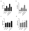
The combination of SL–GAC and IR treatments induced ferroptosis in A549 and H1299 cells. (a,b) The intracellular iron content of A549 (a) and H1299 (b) cells after treatment with SL–GAC and IR was significantly increased compared with that in the control group. (c,d) The level of MDA in the combined treatment group increased, and the combined effect was greater than IR treatment group in A549 (a) and H1299 (b) cells. Data are reported as mean ± SD. * p < 0.05, ** p < 0.01, *** p < 0.001 versus control group; # p < 0.05, ## p < 0.01, ### p < 0.001 versus SL–GAC+IR groups. Data are representative from three parallel experiments.
Figure 6
Figure 6
Fer-1 rescued ferroptosis induced by SL–GAC treatment and the resulting radio sensitization in A549 and H1299 cells. (ac) Fer-1 rescues the SL–GAC-induced proliferation inhibition in A549 and H1299 cells. (d) Fer-1 rescued radio sensitization caused by SL–GAC in A549 and H1299 cells in clone formation experiments. (e) Fer-1 modulated the expression levels of iron death-related proteins in A549 and H1299 cells treated with SL–GAC and IR groups. ** p < 0.01, *** p < 0.001 versus control group; ## p < 0.01, ### p < 0.001 versus SL–GAC+IR groups. Data are representative from three parallel experiments.

References

    1. Sung H., Ferlay J., Siegel R.L., Laversanne M., Soerjomataram I., Jemal A., Bray F. Global Cancer Statistics 2020: GLOBOCAN Estimates of Incidence and Mortality Worldwide for 36 Cancers in 185 Countries. Cancer J. Clin. 2021;71:209–249. doi: 10.3322/caac.21660. - DOI - PubMed
    1. Herbst R.S., Morgensztern D., Boshoff C. The biology and management of non-small cell lung cancer. Nature. 2018;553:446–454. doi: 10.1038/nature25183. - DOI - PubMed
    1. Li Y., Yan B., He S. Advances and challenges in the treatment of lung cancer. Biomed. Pharmacother. 2023;169:115891. - PubMed
    1. Baatout S., editor. Radiobiology Textbook. Springer International Publishing; Cham, Switzerland: 2023. pp. 237–238. - DOI
    1. Wu Y., Song Y., Wang R., Wang T. Molecular mechanisms of tumor resistance to radiotherapy. Mol. Cancer. 2023;22:96. doi: 10.1186/s12943-023-01801-2. - DOI - PMC - PubMed

MeSH terms

LinkOut - more resources