Review
. 2025 Mar 25;14(7):1027.
doi: 10.3390/plants14071027.

The Extended Synaptotagmins of Physcomitrium patens

Affiliations
Review

The Extended Synaptotagmins of Physcomitrium patens

Alexander Kaier et al. Plants (Basel). .

Abstract

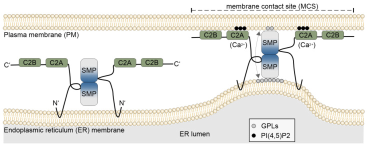
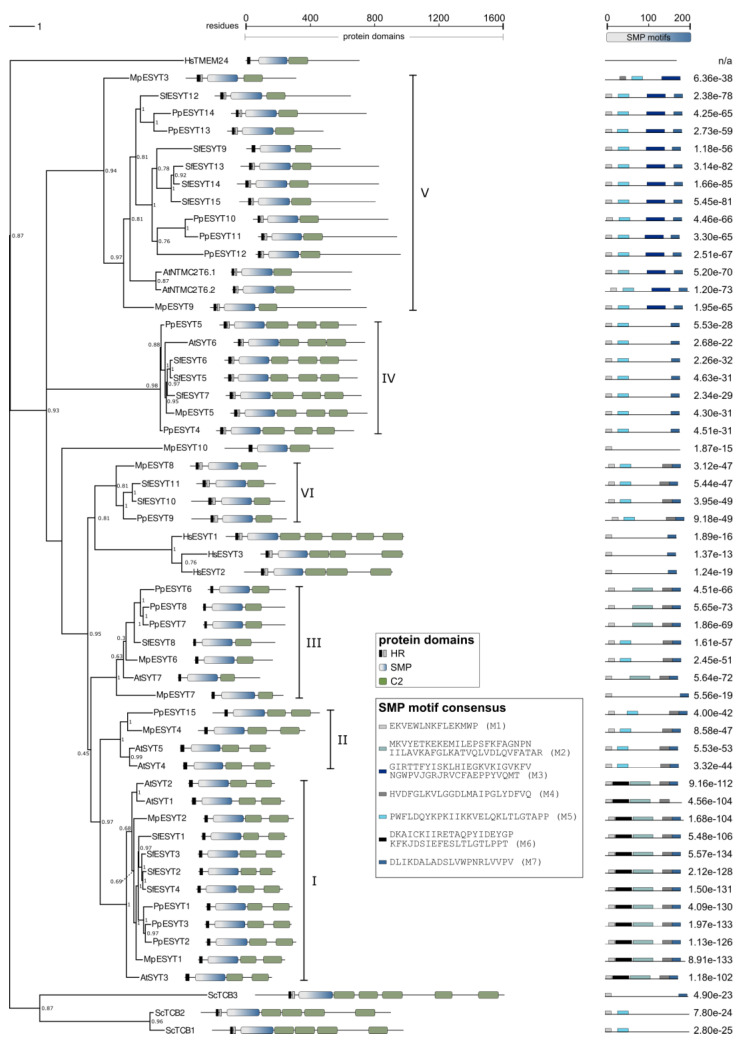
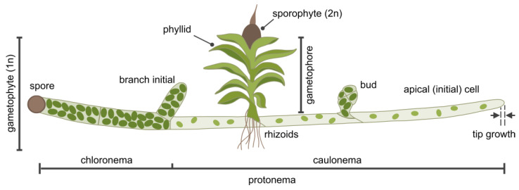
Membrane contact sites (MCSs) between the endoplasmic reticulum and the plasma membrane enable the transport of lipids without membrane fusion. Extended Synaptotagmins (ESYTs) act at MCSs, functioning as tethers between two membrane compartments. In plants, ESYTs have been mainly investigated in A. thaliana and shown to maintain the integrity of the plasma membrane, especially during stress responses like cold acclimatization, mechanical trauma, and salt stress. ESYTs are present at the MCSs of plasmodesmata, where they regulate defense responses by modulating cell-to-cell transfer of pathogens. Here, the analysis of ESYTs was expanded to the bryophyte Physcomitrium patens, an extant representative of the earliest land plant lineages. P. patens was found to contain a large number of ESYTs, distributed over all previously established classes and an additional class not present in A. thaliana. Motif discovery identified regions in the Synaptotagmin-like mitochondrial (SMP) domain that may explain phylogenetic relationships as well as protein function. The adaptation mechanisms of P. patens necessary to conquer land and its simple tissue structure make it highly suitable as a model organism to study ESYT functions in tip growth, stress responses, and plasmodesmata-mediated transport, and open new directions of research regarding the function of MCSs in cellular processes and plant evolution.

Keywords: Physcomitrium patens; bryophytes; extended synaptotagmins; lipid transport proteins; membrane contact sites; plasmodesmata; tip growth.


Conflict of interest statement

The authors declare no conflicts of interest.

Figures

Figure 1
Figure 1
Schematic drawing of Extended Synaptotagmins. ESYT dimers are anchored by their N-terminus to the endoplasmic reticulum (ER), while Ca2+-dependent membrane-targeting domains (C2: green box) at the C-terminus can bind to the plasma membrane (PM), depending on the presence of Ca2+ ions and the local lipid composition of the PM [54]. Attachment of ESYTs to the PM forms tethers that reduce the distance between the two membrane compartments, creating a membrane contact site (MCS). The two Synaptotagmin-like Mitochondrial-lipid-binding Protein (SMP: gray–blue gradient box) domains form a dimer by interacting in an antiparallel configuration at their respective C-terminal end and associating at their N-terminal regions with the ER and the PM, allowing the transfer of lipids between the ER and the PM, causing changes in the lipid composition at the MCS. One such example is the transfer of glycerophospholipids (GPLs), e.g., phosphatidylethanolamine, phosphatidylcholine, and phosphatidylserine from the ER to the PM, but ESYTs can exchange lipids in both directions.
Figure 3
Figure 3
Phylogenetic tree of ESYT homologs. ESYT domain structure and overrepresented SMP motifs are shown. Protein sequences of ESYT homologs and HsTMEM24 of H. sapiens [48,68], S. cerevisiae [56], and A. thaliana [5,22,72] were reported previously. ESYT homologs of P. patens, S. phallax, and M. polymorpha were identified using tBLASTn [125] (protein query against translated nucleotide databases) with genome databases deposited at Phytozome (v13) [126]. The phylogenetic tree was constructed by maximum likelihood and 1000 bootstrap iterations using the phangorn R package [127] (see Supplementary Code S1.R for details). Bootstrap values are shown for the nodes. The branch length bar (upper left scale) represents the average number of substitutions per residue. For a tree containing branch lengths, see Figure S2. The tree was edited using Inkscape [128]. Protein domains were identified using InterPro [129] and SMART [130] analyses. HR: hydrophobic region (black box, single HR; black and gray boxes, hairpin structure), SMP: Synaptotagmin-like Mitochondrial-lipid-binding Protein (gray–blue gradient box), C2: Ca 2+-dependent membrane-targeting domain (green box). SMP motifs M1–M7 (shades of gray and blue boxes) were identified using MEME [124], and the corresponding consensus sequences are displayed. Numbers adjacent to motifs depict the combined match p-value derived from the product of position p-values of the MEME output. Scales indicate amino acid residues of full-length sequences (upper middle scale) or SMP domains (upper right scale). HsTMEM24 could not be aligned in BLASTP or produce motifs in MEME analysis due to low homology to the other sequences; n/a: not applicable.
Figure 2
Figure 2
Schematic depiction of P. patens development. The haploid gametophyte stage begins with a spore, which, after its first cell division, gives rise to chloronema cells containing numerous large chloroplasts and perpendicular cell walls between neighboring cells. Chloronemata differentiate into caulonemata, which have fewer, smaller chloroplasts and are separated by oblique cell walls. Together, chloronema and caulonema cells form the protonema tissue. The apical (initial) cell of each filament expands through tip growth and is the only cell that divides. Side branch initial cells form new filaments. These cells may develop into filaments identical to the main filament or into buds that grow into gametophores, composed of leaf-like structures (phyllids) and rhizoids. At the center of the gametophore apical region, male and female reproductive tissues develop [7], leading to the formation of the diploid sporophyte upon fertilization. After maturation, the sporophyte releases haploid spores, completing the life cycle of P. patens. Not to scale.

Similar articles

References

    1. Saheki Y., De Camilli P. Endoplasmic Reticulum-Plasma Membrane Contact Sites. Annu. Rev. Biochem. 2017;86:659–684. doi: 10.1146/annurev-biochem-061516-044932. - DOI - PubMed
    1. Toulmay A., Prinz W.A. A conserved membrane-binding domain targets proteins to organelle contact sites. J. Cell Sci. 2012;125:49–58. doi: 10.1242/jcs.085118. - DOI - PMC - PubMed
    1. Giordano F., Saheki Y., Idevall-Hagren O., Colombo S.F., Pirruccello M., Milosevic I., Gracheva E.O., Bagriantsev S.N., Borgese N., De Camilli P. PI(4,5)P2-Dependent and Ca2+-Regulated ER-PM Interactions Mediated by the Extended Synaptotagmins. Cell. 2013;153:1494–1509. doi: 10.1016/j.cell.2013.05.026. - DOI - PMC - PubMed
    1. Saheki Y., De Camilli P. The Extended-Synaptotagmins. Biochim. Et Biophys. Acta (BBA)-Mol. Cell Res. 2017;1864:1490–1493. doi: 10.1016/j.bbamcr.2017.03.013. - DOI - PMC - PubMed
    1. Schapire A.L., Voigt B., Jasik J., Rosado A., Lopez-Cobollo R., Menzel D., Salinas J., Mancuso S., Valpuesta V., Baluska F., et al. Arabidopsis synaptotagmin 1 is required for the maintenance of plasma membrane integrity and cell viability. Plant Cell. 2008;20:3374–3388. doi: 10.1105/tpc.108.063859. - DOI - PMC - PubMed

LinkOut - more resources