. 2025 Apr 12:271678X251333256.
doi: 10.1177/0271678X251333256. Online ahead of print.

Impact of hypertension on cerebral small vessel disease: A post-mortem study of microvascular pathology from normal-appearing white matter into white matter hyperintensities

Affiliations

Impact of hypertension on cerebral small vessel disease: A post-mortem study of microvascular pathology from normal-appearing white matter into white matter hyperintensities

Gemma Solé-Guardia et al. J Cereb Blood Flow Metab. .

Abstract

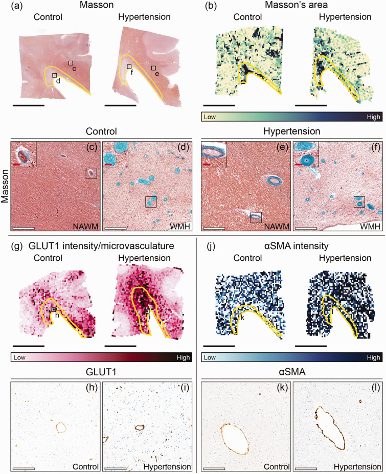
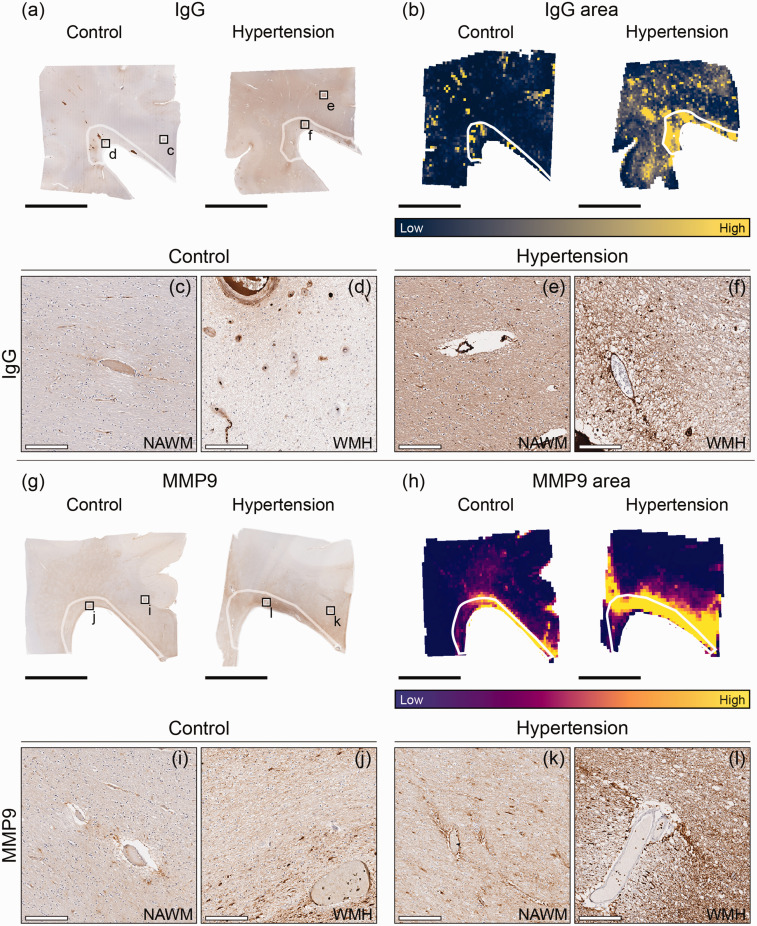
Cerebral small vessel disease (SVD) is diagnosed through imaging hallmarks like white matter hyperintensities (WMH). Novel hypotheses imply that endothelial dysfunction, blood-brain barrier (BBB) disruption and neurovascular inflammation contribute to conversion of normal-appearing white matter (NAWM) into WMH in hypertensive individuals. Aiming to unravel the association between chronic hypertension and the earliest WMH pathogenesis, we characterized microvascular pathology in periventricular NAWM into WMH in post-mortem brains of individuals with and without hypertension. Our second aim was to delineate the NAWM-WMH transition from NAWM towards the center of WMH using deep learning, refining WMH segmentation capturing increases in FLAIR signal. Finally, we aimed to demonstrate whether these processes may synergistically contribute to WMH pathogenesis by performing voxel-wise correlations between MRI and microvascular pathology. Larger endothelium disruption, BBB damage and neurovascular inflammation were observed in individuals with hypertension. We did not observe gradual BBB damage nor neurovascular inflammation along the NAWM-WMH transition. We found a strong correlation between BBB damage and neurovascular inflammation in all individuals in both periventricular NAWM and WMH. These novel findings suggest that endothelium disruption, BBB damage and neurovascular inflammation are major contributors to SVD progression, but being already present in NAWM in hypertension.

Keywords: Blood-brain barrier; MRI; hypertension; immunohistochemistry; inflammation; neuropathology; small vessel disease.


Conflict of interest statement

The author(s) declared no potential conflicts of interest with respect to the research, authorship, and/or publication of this article.

Figures

Figure 1.
Figure 1.
MRI and microvascular pathology study workflow overview. The left hemisphere was divided into ventral part for MRI scanning. (a) Schematic brain image. The grey square illustrates the dimensions of the ventral part of the left hemisphere. The black square illustrates the biopsy used for microvascular pathology stainings of approximately 2 × 2 × 0.5 cm. Sampling of the periventricular NAWM and WMH was successful for all individuals. (b) Image of corresponding MRI fluid-attenuated inversion recovery (FLAIR) axial slab of region of interest. White matter hyperintensity (WMH) before co-registration is outlined with a yellow dotted line. (c) Landmark-based MRI image registered to haematoxylin/eosin (HE). (d) Biopsy. (e) Histology section of haematoxylin/eosin (HE) and (f) Other immunohistochemistry sections were automatically registered to HE, including Masson’s staining. This figure was partly generated with Biorender.com. (L lateral, R Rostral, V Ventral) (scale bar = 1 cm).
Figure 2.
Figure 2.
Immunohistopathological markers for microvascular pathology. (a) In this study we have included several stainings to examine microvascular pathology including microvascular endothelium, vessel wall and BBB damage, and neurovascular inflammation. (b) Shows close-up region from whole slide images for Masson, αSmooth Muscle Actin (αSMA), glucose transporter 1 (GLUT1), Immunoglobulin G (IgG) and matrix metalloproteinase 9 (MMP9) from left to right. (c) Outcome of color deconvolution for each of these stainings. Grey square represents the region shown in panel E. (d) List of parameters studied from each staining and (e) Representative region from Masson’s staining to illustrate the examination of vessel stenosis index. After color deconvolution and thresholding of the images, vessel stenosis index was automatically calculated by dividing the vessel wall area by their total area (vessel wall + lumen area) in ImageJ (scale bar = 200 µm).
Figure 3.
Figure 3.
Immunohistopathological characterization of microvascular endothelium and vessel wall damage in individuals with hypertension and control individuals throughout periventricular white matter. Representative whole slide images of Masson’s staining of brains of normotensive individuals and individuals with hypertension (a) and their corresponding area heatmaps (b). The black squares (a) indicate regions of interest visible in C–F. The yellow outlines in image B depict white matter hyperintensities (WMH). Close ups (X20 magnification) are placed on the upper left corner of (c–f) and show the vessel wall thickening within WMH. G Representative glucose transporter 1 (GLUT1) intensity per microvasculature heatmaps of brains of normotensive individuals and individuals with hypertension. The black squares (g) indicate regions of interest visible in H,I. J Representative αSmooth Muscle Actin (αSMA) intensity heatmaps of brains of normotensive individuals and individuals with hypertension. The black squares (j) indicate regions of interest visible in k,l (black scale bar = 1 cm; white scale bar = 200 µm; red scale bar = 25 µm).
Figure 4.
Figure 4.
Immunohistopathological characterization of blood-brain barrier damage and perivascular MMP9 in individuals with hypertension and control individuals throughout periventricular white matter. Representative whole slide images of Immunoglobulin G (IgG) of brains of normotensive individuals and individuals with hypertension (a) and their corresponding area heatmaps (b). The black squares (a) indicate regions of interest visible in c–f. The white outlines in images B,H depict white matter hyperintensities (WMH). Representative whole slide images of matrix metalloproteinase 9 (MMP9) of brains of normotensive individuals and individuals with hypertension (g) and their corresponding area heatmaps (h). The black squares (g) indicate regions of interest visible in I–L (black scale bar = 1 cm; white scale bar = 200 µm).

Similar articles

References

    1. Wardlaw JM, Smith C, Dichgans M. Small vessel disease: mechanisms and clinical implications. Lancet Neurol 2019; 18: 684–696. - PubMed
    1. Hilal S, Mok V, Youn YC, et al.. Prevalence, risk factors and consequences of cerebral small vessel diseases: data from three Asian countries. J Neurol Neurosurg Psychiatry 2017; 88: 669–674. - PubMed
    1. Pantoni L. Cerebral small vessel disease from pathogenesis and clinical characteristics to therapeutic challenges. Lancet Neurol 2010; 9: 689–701. - PubMed
    1. Wardlaw JM, Smith C, Dichgans M. Mechanisms of sporadic cerebral small vessel disease: insights from neuroimaging. Lancet Neurol 2013; 12: 483–497. - PMC - PubMed
    1. Cai M, Jacob MA, van Loenen MR, et al.. Determinants and temporal dynamics of cerebral small vessel disease: 14-Year Follow-Up. Stroke 2022; 53: 2789–2798. - PMC - PubMed

LinkOut - more resources