. 2025 Apr 12;82(1):157.
doi: 10.1007/s00018-025-05673-1.

m6A reader IGF2BP1 facilitates macrophage glycolytic metabolism and fibrotic phenotype by stabilizing THBS1 mRNA to promote pulmonary fibrosis

Affiliations

m6A reader IGF2BP1 facilitates macrophage glycolytic metabolism and fibrotic phenotype by stabilizing THBS1 mRNA to promote pulmonary fibrosis

Yan Hu et al. Cell Mol Life Sci. .

Abstract

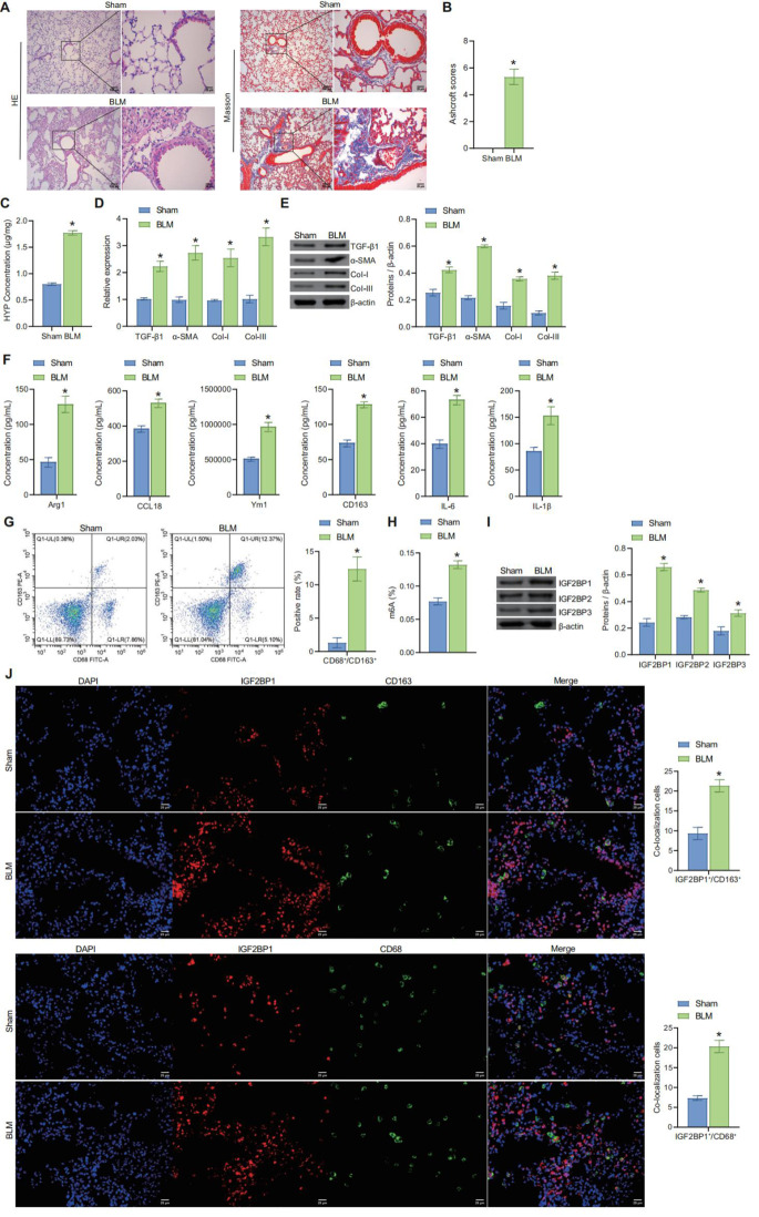
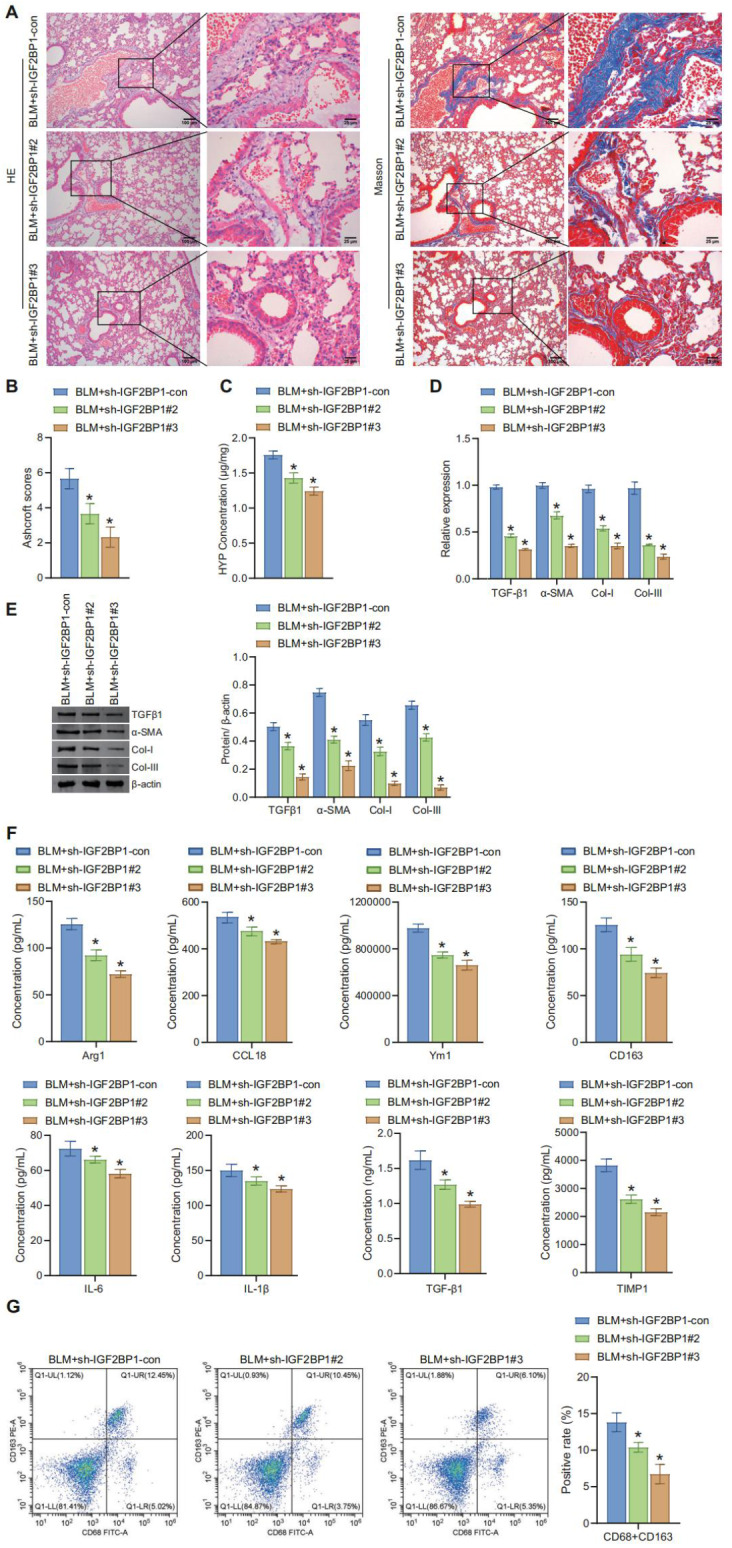
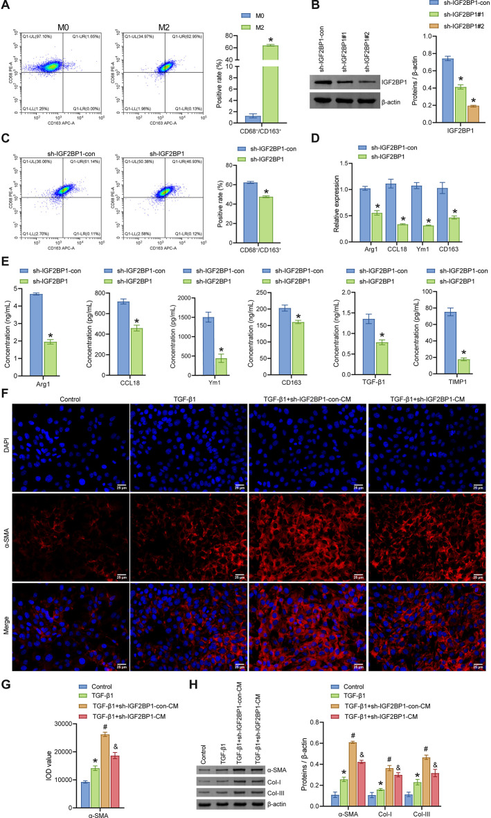
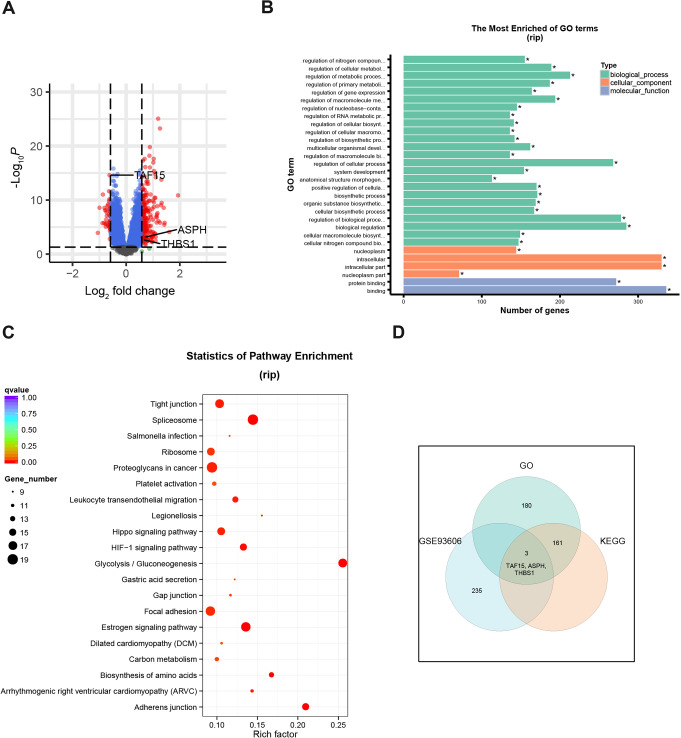
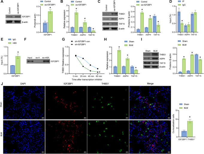
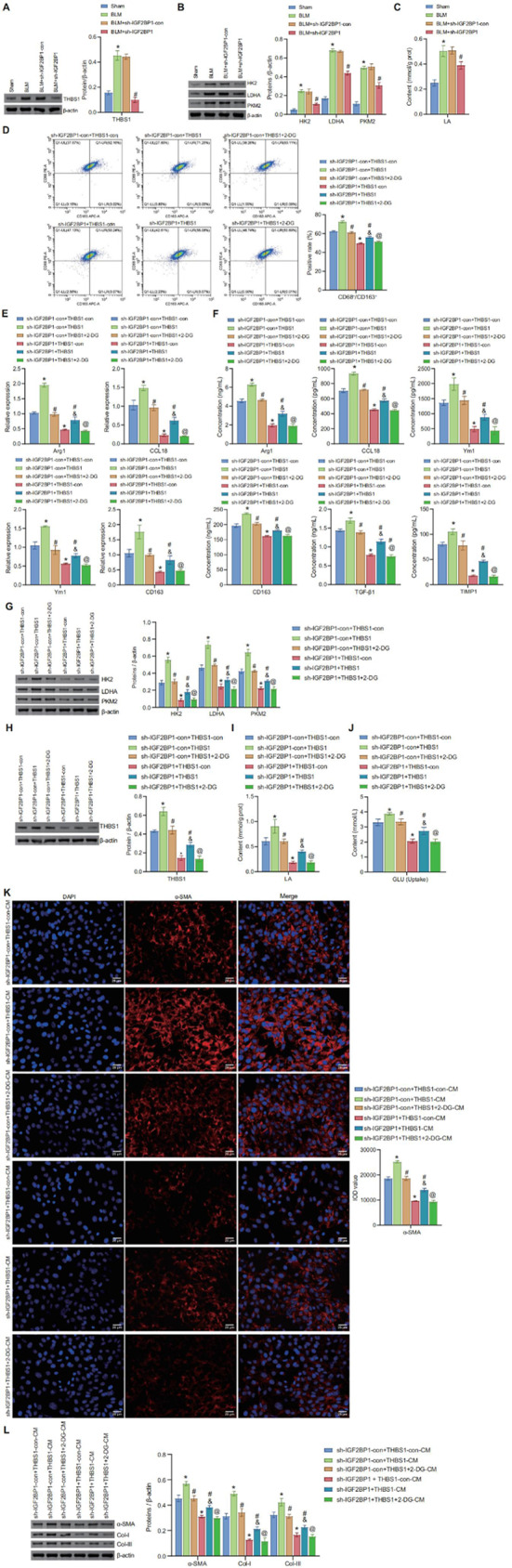
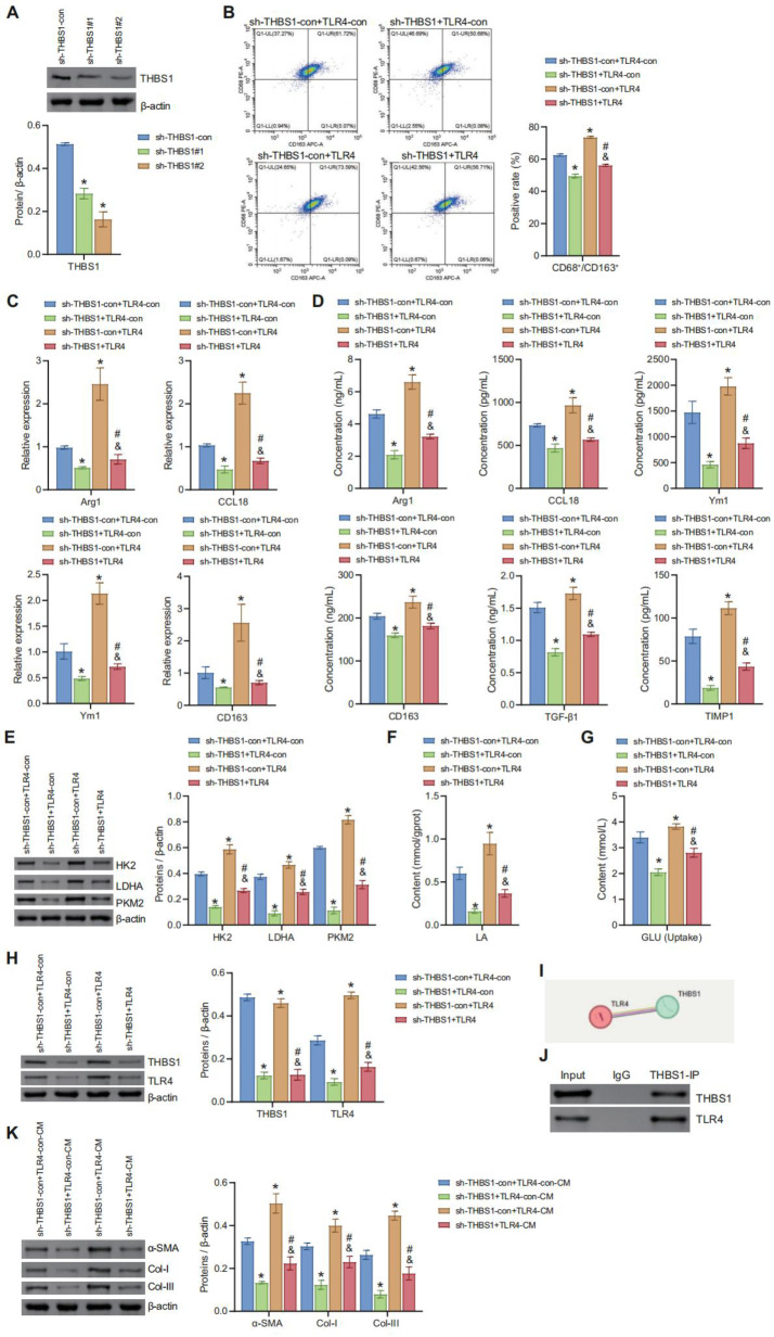
N6-methyladenosine (m6A) modification, a dynamically reversible epigenetic mechanism, is implicated in pulmonary fibrosis (PF) progression. The function and molecular mechanisms of m6A reader, insulin-like growth factor 2 mRNA binding protein 1 (IGF2BP1) in PF remain elusive. This study investigates the mechanistic contributions of IGF2BP1 to PF development. We found IGF2BP1 was overexpressed in macrophages of PF mice. IGF2BP1 knockdown markedly attenuated bleomycin (BLM)-induced lung pathology, as evidenced by reduced inflammatory cell infiltration, fibroblast accumulation, Ashcroft fibrosis scores, and hydroxyproline deposition. Furthermore, IGF2BP1 knockdown downregulated PF-associated markers in lung tissues and embryonic lung fibroblasts (ELFs), including TGF-β1, α-SMA, Collagen-I/III, Arg1, CCL18, Ym1, CD163, IL-6, IL-1β, and TIMP1, while decreasing the CD68+/CD163+ macrophage proportion. Mechanistic studies revealed that IGF2BP1 bound to and stabilized thrombospondin-1 (THBS1) in an m6A-dependent manner. THBS1 overexpression rescued the suppression of macrophage M2 polarization caused by IGF2BP1 knockdown. Additionally, THBS1 overexpression counteracted IGF2BP1 knockdown-mediated inhibition of glycolysis, restoring HK2, LDHA, and PKM2 expression, lactate/glucose metabolism, and ATP production. Intriguingly, THBS1 physically interacted with toll-like receptor 4 (TLR4), and TLR4 overexpression reversed the inhibitory effect of THBS1 knockdown on macrophage M2 polarization and glycolytic reprogramming. Collectively, our findings demonstrate that IGF2BP1 drives PF progression by stabilizing THBS1 mRNA via m6A modification, thereby promoting TLR4-mediated macrophage M2 polarization and glycolytic activation. This study unveils a novel IGF2BP1/THBS1/TLR4 regulatory axis in PF pathogenesis, offering potential therapeutic targets.

Keywords: Glycolytic metabolism; Insulin-like growth factor 2 mRNA binding protein 1; Macrophage polarization; Pulmonary fibrosis; Thrombospondin-1; n6-methyladenosine.


Conflict of interest statement

Declarations. Ethics approval: Animal ethical approval for the present study was obtained from the Institutional Animal Care and Use Committee, The Second Xiangya Hospital of Central South University, China (Approval number: 202400044). Consent to participate: N/A. Consent for publish: N/A. Competing interests: All authors declare no conflicts of interest associated with this manuscript.

Figures

Fig. 1
Fig. 1
Increased m6A modification during fibrosis process, with overexpression of IGF2BP1 in macrophages of PF mice (A) Morphological changes in mouse lung tissue were observed through HE staining and Masson’s trichrome staining. Scale bar = 100 μm and 25 μm. (B) Assessment of lung fibrosis severity in mice using Ashcroft score. (C) Measurement of HYP concentration in mouse lung tissue using a biochemical assay kit. (D) and (E) Expression of TGF-β1, α-SMA, Col-I, and Col-III at the mRNA and protein levels in mouse lung tissue was analyzed by qRT-PCR and WB, respectively. (F) Levels of Arg1, CCL18, Ym1, CD163, IL-6, and IL-1β in the BALF were detected using a biochemical assay kit. (G) Flow cytometry analysis of M2 macrophage infiltration in mouse lung tissue. (H) Evaluation of total RNA m6A modification levels in mouse lung tissue using colorimetric assay. (I) Protein expression of IGF2BP1, IGF2BP2, and IGF2BP3 in mouse lung tissue was detected by WB. (J) Co-expression analysis of IGF2BP1 and CD163, as well as IGF2BP1 and CD68, in macrophages using IF double-staining. Scale bar = 25 μm. *P < 0.05 vs. Sham
Fig. 2
Fig. 2
Knockdown of IGF2BP1 suppresses fibrosis and M2 macrophage infiltration in PF mice (A) Morphological changes in mouse lung tissue were observed through HE staining and Masson’s trichrome staining. Scale bar = 100 μm and 25 μm. (B) Assessment of lung fibrosis severity in mice using Ashcroft score. (C) Measurement of HYP concentration in mouse lung tissue using a biochemical assay kit. (D) and (E) Expression of TGF-β1, α-SMA, Col-I, and Col-III at the mRNA and protein levels in mouse lung tissue was analyzed by qRT-PCR and WB, respectively. (F) Levels of Arg1, CCL18, Ym1, CD163, IL-6, IL-1β, TGF-β1, and TIMP1 in the BALF were detected using biochemical assays. (G) Flow cytometry analysis of M2 macrophage infiltration in mouse lung tissue. *P < 0.05 vs. BLM + sh-IGF2BP1-con
Fig. 3
Fig. 3
Knockdown of IGF2BP1 inhibits macrophage M2 polarization and alleviates fibrosis (A) Flow cytometry analysis of M2 polarization in BMDMs. *P < 0.05 vs. M0. (B) Knockdown efficiency of sh-IGF2BP1#1 and sh-IGF2BP1#2 was detected by WB. *P < 0.05 vs. sh-IGF2BP1-con. (C) Flow cytometry analysis of M2 polarization in BMDMs. (D) and (E) Levels of Arg1, CCL18, Ym1, CD163, TGF-β1, and TIMP1 in the conditioned medium of BMDMs were detected by qRT-PCR and biochemical assays, respectively. *P < 0.05 vs. sh-IGF2BP1-con. (F) and (G) IF staining to detect α-SMA expression in ELFs. Scale bar = 25 μm. (H) Protein expression of α-SMA, Col-I, and Col-III in ELFs was detected by WB. *P < 0.05 vs. Control, #P < 0.05 vs. TGF-β1, &P < 0.05 vs. TGF-β1 + sh-IGF2BP1-CM
Fig. 4
Fig. 4
Identification of potential downstream targets of IGF2BP1 (A) A volcano plot visualizes the expression of 238 DEGs between PF patients and controls (|log2FC| > 0.5, FDR-corrected P < 0.05). Red indicates high expression, blue indicates low. (B) Top 30 significantly enriched GO terms. (C) Top 20 significantly enriched KEGG pathways. (D) Venn diagram depicting the overlap among gene sets
Fig. 5
Fig. 5
IGF2BP1 enhances the stability of THBS1 mRNA in Macrophages (A) Protein expression of IGF2BP1 in BMDMs was detected by WB. (B) and (C) mRNA and protein expression of THBS1, ASPH, and TAF15 in BMDMs analyzed by qRT-PCR and WB, respectively. *P < 0.05 vs. Control. (D) RIP assay to detect the interaction between IGF2BP1 protein and THBS1, ASPH, and TAF15 mRNA. (E) MeRIP analysis of m6A methylation levels of THBS1 in BMDMs. *P < 0.05 vs. IgG. (F) The RNA pull-down assay verified that the IGF2BP1 protein selectively binds to the methylated bait of THBS1, rather than the unmethylated control (ss-A). (G) Actinomycin D experiment to assess the stability of THBS1 mRNA. *P < 0.05 vs. sh-IGF2BP1-con. (H) and (I) mRNA and protein expression of THBS1, ASPH, and TAF15 in mouse lung tissue analyzed by qRT-PCR and WB, respectively. (J) Co-expression analysis of THBS1 and IGF2BP1 in mouse lung tissue using IF double-staining. Scale bar = 25 μm. *P < 0.05 vs. Sham
Fig. 6
Fig. 6
IGF2BP1 enhances macrophage M2 polarization through upregulation of THBS1-mediated glycolytic metabolism (A) Protein expression of THBS1 in mouse lung tissue was detected by WB. (B) Protein expression of HK2, LDHA, and PKM2 in mouse lung tissue was detected by WB. (C) Levels of LA production in mouse lung tissue were detected using biochemical assay kits. *P < 0.05 vs. Sham, #P < 0.05 vs. BLM + sh-IGF2BP1-con. (D) Flow cytometry analysis of M2 polarization in BMDMs. (E) and (F) Levels of Arg1, CCL18, Ym1, CD163, TGF-β1, and TIMP1 in BMDMs were detected by qRT-PCR and biochemical assay kits, respectively. (G) Protein expression of HK2, LDHA, and PKM2 in mouse BMDMs was detected by WB. (H) Protein expression of THBS1 in BMDMs was detected by WB. (I) and (J) LA production and GLU uptake in BMDMs were detected using biochemical assays. *P < 0.05 vs. sh-IGF2BP1-con + THBS1-con, #P < 0.05 vs. sh-IGF2BP1-con + THBS1, &P < 0.05 vs. sh-IGF2BP1 + THBS1-con, @P < 0.05 vs. sh-IGF2BP1 + THBS1. (K) IF staining to detect α-SMA expression in ELFs. Scale bar = 25 μm. (L) Protein expression of α-SMA, Col-I, and Col-III in ELFs was detected by WB. *P < 0.05 vs. sh-IGF2BP1-con + THBS1-con-CM, #P < 0.05 vs. sh-IGF2BP1-con + THBS1-CM, &P < 0.05 vs. sh-IGF2BP1 + THBS1-con-CM, @P < 0.05 vs. sh-IGF2BP1 + THBS1-CM
Fig. 7
Fig. 7
THBS1 promotes glycolytic metabolism and macrophage M2 polarization via TLR4 signaling (A) Knockdown efficiency of sh-THBS1#1 and sh-THBS1#2 was detected by WB. *P < 0.05 vs. sh THBS1-con. (B) Flow cytometry analysis of M2 polarization in BMDMs. (C) and (D) Levels of Arg1, CCL18, Ym1, CD163, TGF-β1, and TIMP1 in the conditioned medium of BMDMs were detected by qRT-PCR and biochemical assays, respectively. (E) Protein expression of HK2, LDHA, and PKM2 in mouse BMDMs was detected by WB. (F) and (G) LA production and GLU uptake in BMDMs were detected using biochemical assays. (H) Protein expression of THBS1 and TLR4 in BMDMs was detected by WB. (I) Potential target relationship between THBS1 and TLR4 was predicted by the STRING database. (J) Co-IP to validate the interaction between THBS1 and TLR4. *P < 0.05 vs. sh-THBS1-con + TLR4-con, #P < 0.05 vs. sh-THBS1 + TLR4-con, &P < 0.05 vs. sh-THBS1-con + TLR4. (K) Protein expression of α-SMA, Col-I, and Col-III in ELFs was detected by WB. *P < 0.05 vs. sh-THBS1-con + TLR4-con-CM, #P < 0.05 vs. sh-THBS1 + TLR4-con-CM, &P < 0.05 vs. sh-THBS1-con + TLR4-CM

Similar articles

References

    1. Meyer KC (2017) Pulmonary fibrosis, part I: epidemiology, pathogenesis, and diagnosis. Expert Rev Respir Med 11:343–359. 10.1080/17476348.2017.1312346 - PubMed
    1. León-Román F, Valenzuela C, Molina-Molina M (2022) Idiopathic pulmonary fibrosis. Med Clin (Barc) 159:189–194. 10.1016/j.medcli.2022.02.020 - PubMed
    1. Kishore A, Petrek M (2021) Roles of macrophage polarization and macrophage-Derived MiRNAs in pulmonary fibrosis. Front Immunol 12:678457. 10.3389/fimmu.2021.678457 - PMC - PubMed
    1. Para R, Romero F, George G, Summer R (2019) Metabolic reprogramming as a driver of fibroblast activation in pulmonaryfibrosis. Am J Med Sci 357:394–398. 10.1016/j.amjms.2019.02.003 - PMC - PubMed
    1. Xie N, Tan Z, Banerjee S, Cui H, Ge J, Liu RM, Bernard K, Thannickal VJ, Liu G (2015) Glycolytic reprogramming in myofibroblast differentiation and lung fibrosis. Am J Respir Crit Care Med 192:1462–1474. 10.1164/rccm.201504-0780OC - PMC - PubMed

MeSH terms