

Brain organoids: building higher-order complexity and neural circuitry models
- PMID: 40221251
- PMCID: PMC12276974
- DOI: 10.1016/j.tibtech.2025.02.009
Brain organoids: building higher-order complexity and neural circuitry models
Abstract
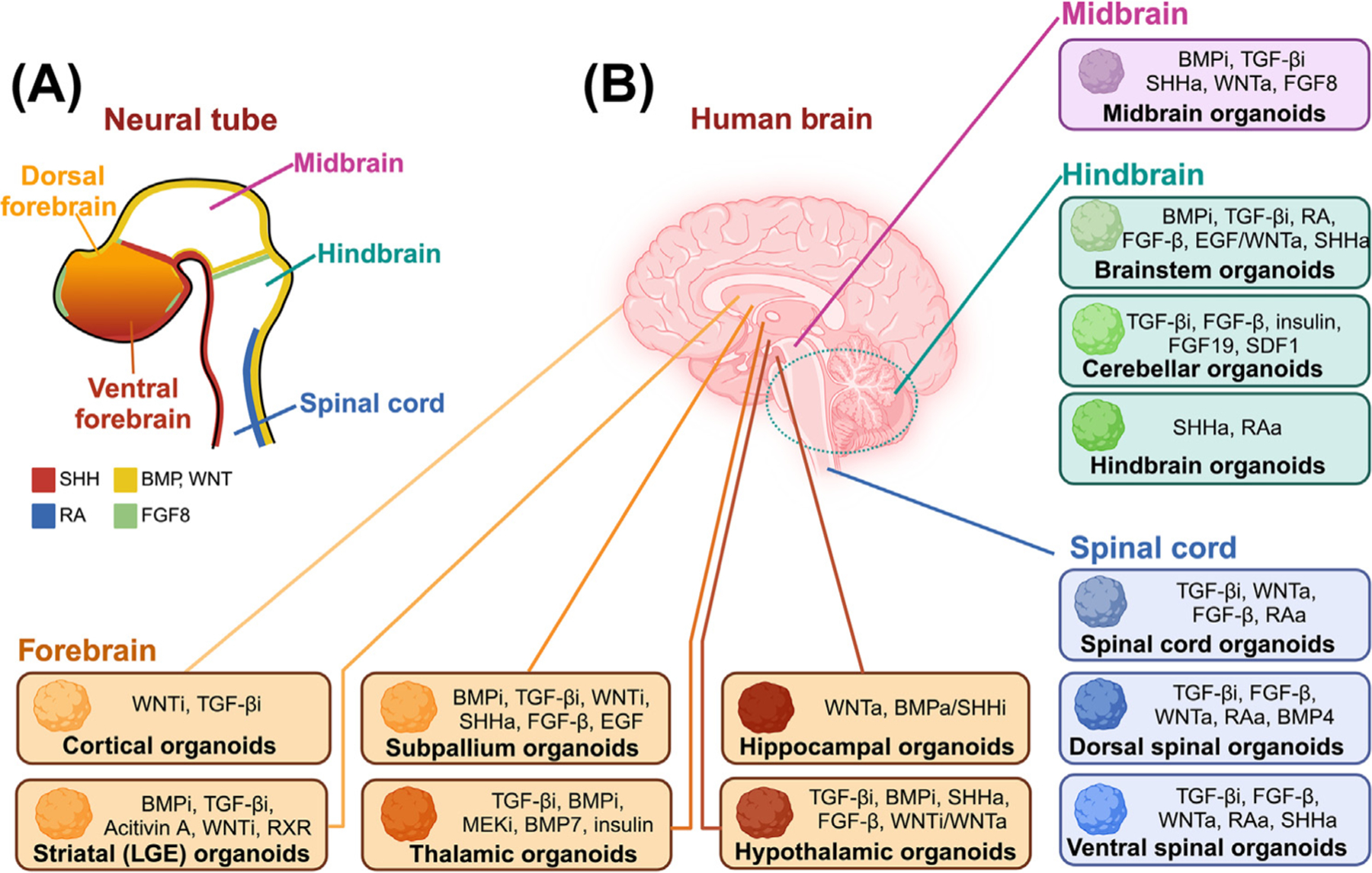
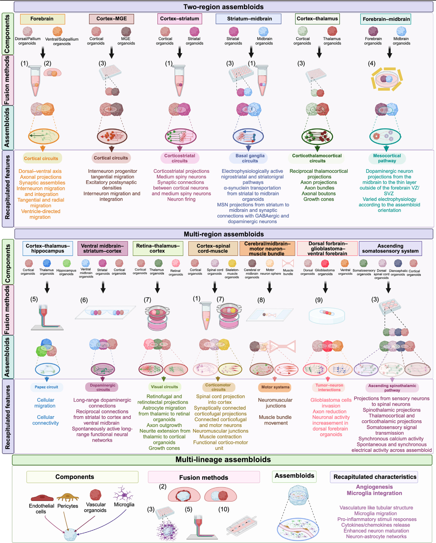
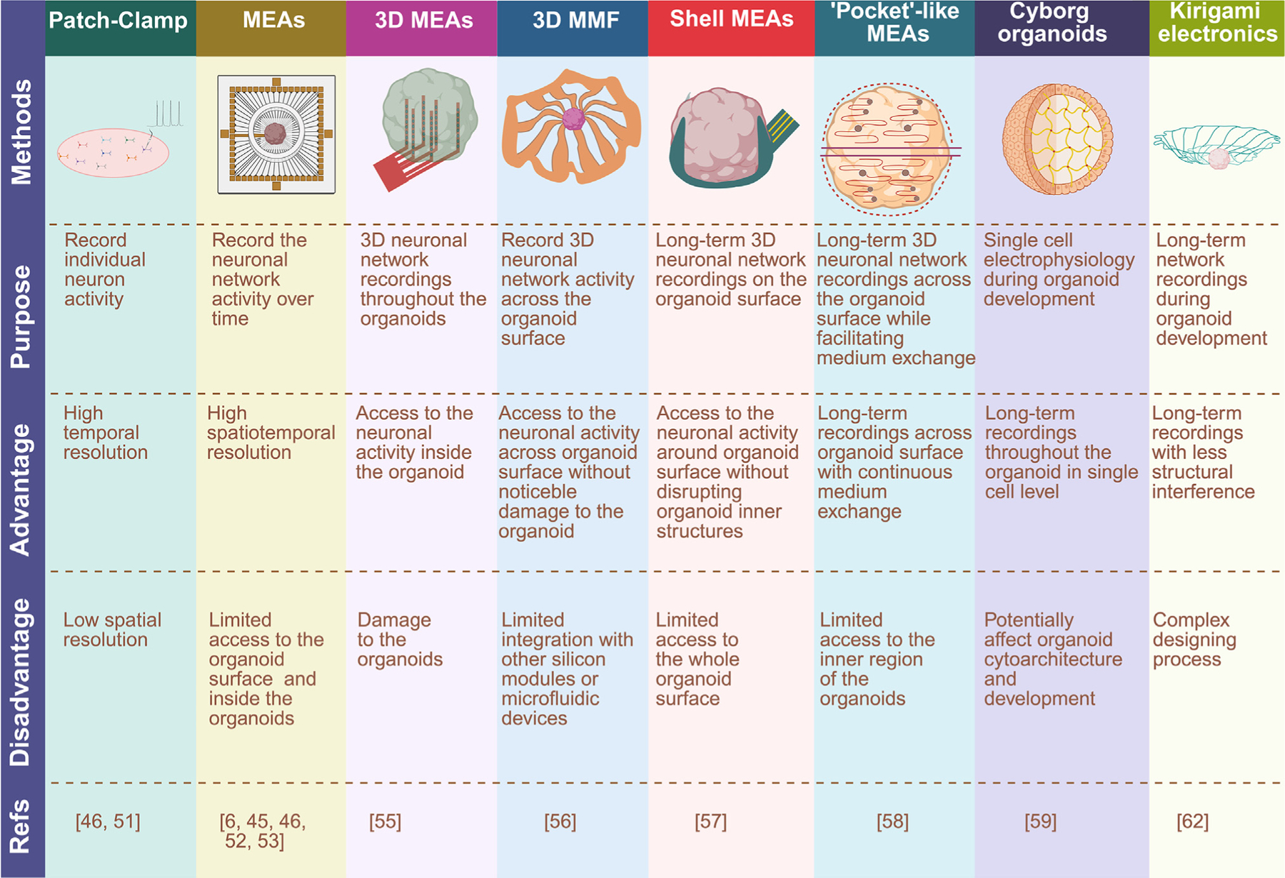
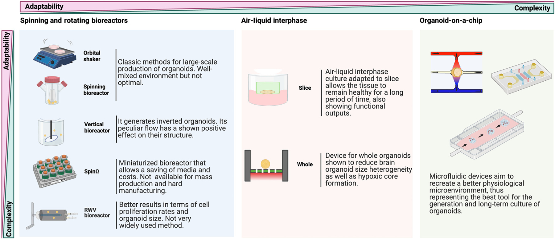
Brain organoids are 3D tissue models of the human brain that are derived from pluripotent stem cells (PSCs). They have enabled studies that were previously stymied by the inaccessibility of human brain tissue or the limitations of mouse models of some brain diseases. Despite their enormous potential, brain organoids have had significant limitations that prevented them from recapitulating the full complexity of the human brain and reduced their utility in disease studies. We describe recent progress in addressing these limitations, especially building complex organoids that recapitulate the interactions between multiple brain regions, and reconstructing in vitro the neural circuitry present in vivo. These major advances in the human brain organoid technology will remarkably facilitate brain disease modeling and neuroscience research.
Keywords: assembloids; brain development; brain disease; brain organoids; neural circuitry.
Copyright © 2025 The Authors. Published by Elsevier Ltd.. All rights reserved.
Conflict of interest statement
Declaration of interests The authors declare no competing interests.
Figures
Similar articles
-
Vascularized Brain Assembloids With Enhanced Cellular Complexity Provide Insights Into the Cellular Deficits of Tauopathy.Stem Cells. 2024 Feb 8;42(2):107-115. doi: 10.1093/stmcls/sxad086. Stem Cells. 2024. PMID: 37995336 Free PMC article.
-
Vascularized Brain Assembloids with Enhanced Cellular Complexity Provide Insights into The Cellular Deficits of Tauopathy.bioRxiv [Preprint]. 2023 Jul 2:2023.06.30.547293. doi: 10.1101/2023.06.30.547293. bioRxiv. 2023. Update in: Stem Cells. 2024 Feb 8;42(2):107-115. doi: 10.1093/stmcls/sxad086. PMID: 37425812 Free PMC article. Updated. Preprint.
-
A Hybrid 2D/3D Approach for Neural Differentiation Into Telencephalic Organoids and Efficient Modulation of FGF8 Signaling.Bio Protoc. 2025 Jun 20;15(12):e5354. doi: 10.21769/BioProtoc.5354. eCollection 2025 Jun 20. Bio Protoc. 2025. PMID: 40620811 Free PMC article.
-
Advances, challenges, and opportunities of human midbrain organoids for modelling of the dopaminergic system.EMBO J. 2025 Aug;44(15):4181-4195. doi: 10.1038/s44318-025-00494-1. Epub 2025 Jul 2. EMBO J. 2025. PMID: 40604323 Free PMC article. Review.
-
How lived experiences of illness trajectories, burdens of treatment, and social inequalities shape service user and caregiver participation in health and social care: a theory-informed qualitative evidence synthesis.Health Soc Care Deliv Res. 2025 Jun;13(24):1-120. doi: 10.3310/HGTQ8159. Health Soc Care Deliv Res. 2025. PMID: 40548558
References
Publication types
MeSH terms
Grants and funding
LinkOut - more resources
Full Text Sources
