. 2025 Apr 12;12(1):615.
doi: 10.1038/s41597-025-04467-1.

TRUSTED: The Paired 3D Transabdominal Ultrasound and CT Human Data for Kidney Segmentation and Registration Research

Affiliations

TRUSTED: The Paired 3D Transabdominal Ultrasound and CT Human Data for Kidney Segmentation and Registration Research

William Ndzimbong et al. Sci Data. .

Abstract

Inter-modal image registration (IMIR) and image segmentation with abdominal Ultrasound (US) data have many important clinical applications, including image-guided surgery, automatic organ measurement, and robotic navigation. However, research is severely limited by the lack of public datasets. We propose TRUSTED (the Tridimensional Renal Ultra Sound TomodEnsitometrie Dataset), comprising paired transabdominal 3DUS and CT kidney images from 48 human patients (96 kidneys), including segmentation, and anatomical landmark annotations by two experienced radiographers. Inter-rater segmentation agreement was over 93% (Dice score), and gold-standard segmentations were generated using the STAPLE algorithm. Seven anatomical landmarks were annotated, for IMIR systems development and evaluation. To validate the dataset's utility, 4 competitive Deep-Learning models for kidney segmentation were benchmarked, yielding average DICE scores from 79.63% to 90.09% for CT, and 70.51% to 80.70% for US images. Four IMIR methods were benchmarked, and Coherent Point Drift performed best with an average Target Registration Error of 4.47 mm and Dice score of 84.10%. The TRUSTED dataset may be used freely to develop and validate segmentation and IMIR methods.


Conflict of interest statement

Competing interests: The authors declare no competing interests.

Figures

Fig. 1
Fig. 1
Visualization of annotated data in the TRUSTED dataset (Patient 418, Annotator 2). Top left: Segmentation of left and right kidneys in CT. Top right: 2DCT slice of the right kidney in the canonical sagittal plane with 7 anatomical landmarks overlaid. Bottom left: Segmentation of the right kidney in 3DUS. Bottom right: 2DUS slice of the right kidney in the canonical sagittal plane with 7 anatomical landmarks overlaid.
Fig. 2
Fig. 2
Qualitative results showing example CT and US segmentations using the TRUSTED dataset (patients 418 and 680). The top two rows show coronal CT slices, with ground-truth segmentations overlaid in blue, and estimated segmentations in green. The bottom two rows show longitudinal US slices, with ground truth segmentations overlaid in red and estimated segmentations in green. The two first columns (Manual Ann.1 and Manual Ann.2) show segmentations from each annotator, and the remaining columns show the best training version on average between single or double target(s) of each automatic segmentation from 4 DNN-based methods.
Fig. 3
Fig. 3
Examples of noisy initial registration (from the best-fitting similarity transform using the noisy landmarks). In the first and the third rows, the checkerboard visualizations show, for different values of σ, the fixed CT image overlaid with the US image after noisy initial registration and the kidney contours. In the second and fourth rows, a 3D visualization of the corresponding masks is shown.
Fig. 4
Fig. 4
Initialization sensitivity trends of registration refinement methods using the TRUSTED dataset. The figure is divided into 4 rows (one row per performance metric). Each row has 6 sub-figures, showing the performance of every compared method using a different amount of artificial noise added to the initial registrations, ranging from std = 0 mm (no noise) to std = 10 mm (large noise). Boxplots are used to summarize the performance distribution of each method in two configurations (using rigid and affine spatial transforms). For each boxplot, the lower, central, and upper bars give the 1st, 2nd (median), and 3rd quartiles. White circles give the mean, and whiskers give the extreme values not considered as outliers. Dots give outlier values, and the numbers above each boxplot give the number of outliers that are beyond the y-axis range.
Fig. 5
Fig. 5
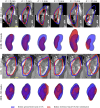
Qualitative results showing registration performance using the TRUSTED dataset. Each figure column corresponds to a method. The first and second figure rows show results with one example (KDY_418R), using a rigid and affine registration refinement transform. The third and fourth figure rows show results with a second example (KDY_680L), using a rigid and affine registration refinement transform. Each image in the figure shows 4 annotations: Blue contour - the GT kidney segmentation in CT. Blue point - the GT position of Landmark 1 in CT. Red contour - the GT kidney segmentation in US, moved to CT coordinates according to the estimated registration. Red point - the GT position of Landmark 1 in US, moved to CT coordinates according to the estimated registration. A checkerboard visualization is used to show the fixed image (CT) overlaid with the registered image (US).

References

    1. ESR Abdominal applications of ultrasound fusion imaging technique: liver, kidney, and pancreas. Insights into Imaging10, 6 (2019). - PMC - PubMed
    1. Leroy, A. et al. Percutaneous renal puncture, requirements and preliminary results. arXiv preprint physics/0610209 (2006).
    1. Fu, Y. et al. Biomechanically constrained non rigid MR-TRUS prostate registration using deep learning based 3d point cloud matching. Medical image analysis67, 101845 (2021). - PMC - PubMed
    1. Arun, K. S., Huang, T. S. & Blostein, S. D. Least-squares fitting of two 3-d point sets. IEEE Transactions on Pattern Analysis and Machine IntelligencePAMI-9, 698–700, 10.1109/TPAMI.1987.4767965 (1987). - PubMed
    1. Agamennoni, G., Fontana, S., Siegwart, R. Y. & Sorrenti, D. G. Point clouds registration with probabilistic data association. In 2016 IEEE/RSJ International Conference on Intelligent Robots and Systems (IROS), 4092–4098, 10.1109/IROS.2016.7759602 (2016).