

Associations between preoperative cerebral white matter microstructural changes and neurodevelopmental deficits in CHD infants: a diffusion tensor imaging study
- PMID: 40221812
- PMCID: PMC11993994
- DOI: 10.1186/s13052-025-01962-4
Associations between preoperative cerebral white matter microstructural changes and neurodevelopmental deficits in CHD infants: a diffusion tensor imaging study
Abstract
Background: Neurodevelopmental deficits(NDs) frequently occur in patients with cyanotic congenital heart disease (CCHD) because of the hemodynamic abnormalities. We aimed to evaluate white matter(WM) microstructural changes in infants with CHD and analyze the relationship between WM microstructural changes and NDs.
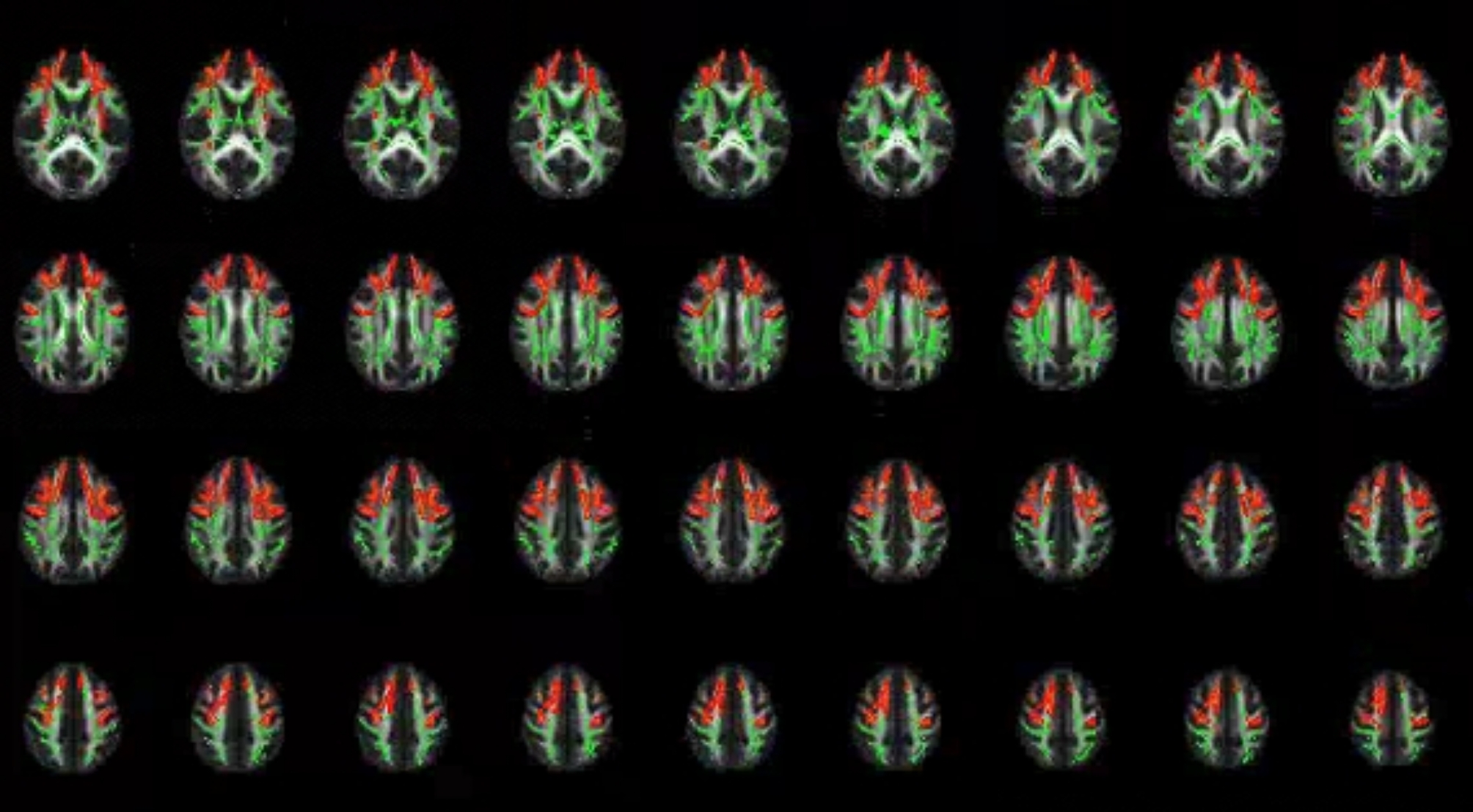
Methods: A total of 40 infants, 20 with CCHD and 20 with ACHD (matched on age and sex), who underwent preoperative DTI scanning were prospectively enrolled in the study. Multiple linear regression analysis were used to investigate the associations between brain microstructural changes and both clinical variables and neurodevelopmental outcomes, assessed with Gesell Developmental Schedules-Third Edition (GDS-III).
Results: Infants with CCHD showed lower fractional anisotropy (FA) values in the bilateral cingulum hippocampus (CGH), right anterior thalamic radiation (ATR), and forceps minor (fminor) and exhibited poorer performance in adaptive, motor, language, and personal-social behaviors (all P < 0.05). For CHD infants, the FA values of fminor were positively correlated with adaptive, fine motor, and language behaviors (P = 0.026, 0.040, and 0.038, respectively). The microstructures of right ATR were positively correlated with adaptive and fine motor behaviors (P = 0.047 and 0.035, respectively), and FA values of right CGH were positively correlated with language behavior (P = 0.007). Hypoxia-related indicators and the internal diameters of the heart and large vessels were associated with neurodevelopmental and brain microstructural changes.
Conclusions: This study suggests that cerebral white matter microstructural changes may serve as imaging markers of neurodevelopmental deficits, with oxygen supply playing a crucial role in white matter microstructural development.
Keywords: Diffusion tensor imaging; Neurodevelopmental deficit; White matter microstructural changes.
© 2025. The Author(s).
Conflict of interest statement
Declarations. Ethics approval and consent to participate: The studies involving human participants were reviewed and approved by the Ethics Committee of Children’s Hospital of Nanjing Medical University. The study is in accordance with the 1964 Helsinki Declaration and its later amendments. Written informed consent to participate in this study was provided by the legal guardian/next of kin of the participants for the publication of any potentially identifiable radiographic images or data included in this article. Consent for publication: Not applicable. Competing interests: The authors have no conflicts of interest to declare.
Figures

Similar articles
-
Diffusion tensor imaging study of pediatric patients with congenital hydrocephalus: 1-year postsurgical outcomes.J Neurosurg Pediatr. 2016 Sep;18(3):306-19. doi: 10.3171/2016.2.PEDS15628. Epub 2016 May 20. J Neurosurg Pediatr. 2016. PMID: 27203134 Free PMC article.
-
Diffusion Tensor MRI of White Matter of Healthy Full-term Newborns: Relationship to Neurodevelopmental Outcomes.Radiology. 2019 Jul;292(1):179-187. doi: 10.1148/radiol.2019182564. Epub 2019 Jun 4. Radiology. 2019. PMID: 31161971 Free PMC article.
-
The effects of mild germinal matrix-intraventricular haemorrhage on the developmental white matter microstructure of preterm neonates: a DTI study.Eur Radiol. 2018 Mar;28(3):1157-1166. doi: 10.1007/s00330-017-5060-0. Epub 2017 Sep 27. Eur Radiol. 2018. PMID: 28956133
-
Brain microstructural development in neonates with critical congenital heart disease: An atlas-based diffusion tensor imaging study.Neuroimage Clin. 2019;21:101672. doi: 10.1016/j.nicl.2019.101672. Epub 2019 Jan 7. Neuroimage Clin. 2019. PMID: 30677732 Free PMC article.
-
The role of diffusion tensor imaging and fractional anisotropy in the evaluation of patients with idiopathic normal pressure hydrocephalus: a literature review.Neurosurg Focus. 2016 Sep;41(3):E12. doi: 10.3171/2016.6.FOCUS16192. Neurosurg Focus. 2016. PMID: 27581308 Review.
References
-
- Bouma BJ, Mulder BJ. Changing landscape of congenital heart disease. Circ Res. 2017;120(6):908–22. - PubMed
-
- van der Bom T, Zomer AC, Zwinderman AH, Meijboom FJ, Bouma BJ, Mulder BJ. The changing epidemiology of congenital heart disease. Nat Rev Cardiol. 2011;8(1):50–60. - PubMed
-
- Mo X, Cai W, Qi J, et al. Expert consensus on nutritional support for children with congenital heart disease (2023 edition). Congeni Heart Dis. 2023;18(6):571–93.
-
- Vida V. Innovations in pediatric and congenital cardiac surgery. Congeni Heart Dis. 2022;17(1):1–3.
MeSH terms
Grants and funding
- 82270310/National Natural Science Foundation of China
- 81970265/National Natural Science Foundation of China
- QDJJ2024003/the postdoctoral start-up funds of Children's Hospital of Nanjing Medical University
- QDJJ2022006/the postdoctoral start-up funds of Children's Hospital of Nanjing Medical University
- QDJJ2023002/the postdoctoral start-up funds of Children's Hospital of Nanjing Medical University
LinkOut - more resources
Full Text Sources
Medical
Miscellaneous
