

Longitudinal MRI in comparison to low-dose CT for follow-up of incidental pulmonary nodules in patients with COPD-a nationwide multicenter trial
- PMID: 40221941
- PMCID: PMC12417304
- DOI: 10.1007/s00330-025-11567-4
Longitudinal MRI in comparison to low-dose CT for follow-up of incidental pulmonary nodules in patients with COPD-a nationwide multicenter trial
Abstract
Purpose: This multicenter trial was conducted to evaluate MRI for the longitudinal management of incidental pulmonary nodules in heavy smokers.
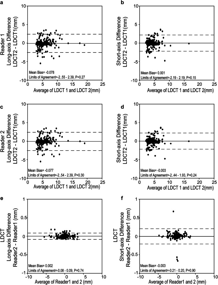
Materials and methods: 239 participants (63.9 ± 8.4 years, 43-82 years) at risk of or with COPD GOLDI-IV from 16 centers prospectively underwent two rounds of same-day low-dose computed tomography (LDCT1&2) and MRI1&2 at an interval of three years in the nationwide COSYCONET trial. All exams were independently assessed for incidental pulmonary nodules in a standardized fashion by two blinded readers, incl. axis measurements and Lung-RADS categorization, with consensual LDCT results serving as the standard of reference. A change in diameter ≥ 2 mm was rated as progress. 11 patients underwent surgery for suspicious nodules after the first round.
Results: Two hundred twenty-four of two hundred forty nodules (93.3%) persisted from LDCT1 to LDCT2, with a sensitivity of MRI2 of 82.8% and 81.5% for readers 1 and 2, respectively. Agreement in Lung-RADS categories between LDCT2 and MRI2 was substantial in per-nodule (κ = 0.62-0.70) and excellent in a per-patient (κ = 0.86-0.88) approach for both readers, respectively. Concordance between LDCT2 and MRI2 for growth was excellent to almost perfect (κ = 0.88-1.0). The accuracy of LDCT1 and MRI1 for lung cancer was 87.5%. Lung-RADS ≥ 3 category on MRI1 had higher accuracy for predicting progress (23.1% and 21.4%, respectively) than LDCT1 (15.8%).
Conclusion: Compared to LDCT, MRI shows similar capabilities for the longitudinal evaluation of incidental nodules in heavy smokers. Decision-making for nodule management guided by Lung-RADS seems feasible based on longitudinal MRI.
Key points: Question Can MRI serve as an alternative to low-dose CT (LDCT) for the longitudinal management of pulmonary nodules in heavy smokers, addressing concerns over radiation exposure? Findings MRI demonstrated substantial agreement with LDCT in detecting nodule growth, accurately categorizing Lung-RADS, and comparable accuracy in identifying malignancy over a three-year follow-up. Clinical relevance Longitudinal MRI demonstrates high consistency with LDCT in assessing the growth of incidental pulmonary nodules and categorizing per-patient Lung-RADS, offering a reliable, radiation-free alternative for monitoring and early malignancy detection in high-risk populations.
Keywords: Chronic obstructive pulmonary disease; Longitudinal management; Low-dose computed tomography; Magnetic resonance imaging; Pulmonary nodules.
© 2025. The Author(s).
Conflict of interest statement
Compliance with ethical standards. Guarantor: The scientific guarantor of this publication is Mark O. Wielpütz. Conflict of interest: The authors of this manuscript declare no relationships with any companies, whose products or services may be related to the subject matter of the article. Statistics and biometry: No complex statistical methods were necessary for this paper. Informed consent: Written informed consent was obtained from all patients in this study. Ethical approval: Institutional Review Board approval was obtained. Study subjects or cohorts overlap: Some study subjects have been previously reported in our previous first-round research [17]. Methodology: Prospective Diagnostic or prognostic study Multicenter study
Figures


References
-
- Maldonado F, Bartholmai BJ, Swensen SJ, Midthun DE, Decker PA, Jett JR (2010) Are airflow obstruction and radiographic evidence of emphysema risk factors for lung cancer? A nested case-control study using quantitative emphysema analysis. Chest 138:1295–1302 - PubMed
-
- Turner MC, Chen Y, Krewski D, Calle EE, Thun MJ (2007) Chronic obstructive pulmonary disease is associated with lung cancer mortality in a prospective study of never smokers. Am J Respir Crit Care Med 176:285–290 - PubMed
-
- Dawson Q (2020) NELSON trial: reduced lung-cancer mortality with volume CT screening. Lancet Respir Med 8:236 - PubMed
-
- Sodickson A, Baeyens PF, Andriole KP et al (2009) Recurrent CT, cumulative radiation exposure, and associated radiation-induced cancer risks from CT of adults. Radiology 251:175–184 - PubMed
Publication types
MeSH terms
Grants and funding
- 82DZL004A1/German Federal Ministry of Education and Research BMBF to the German Center for Lung research
- 82DZL009B1/German Federal Ministry of Education and Research BMBF to the German Center for Lung research
- 22PJD069/Shanghai Pujiang Program
- 2022YQ060/d Shanghai Health Research Foundation for Talents
- 82071873/National Natural Science Foundation of China
LinkOut - more resources
Full Text Sources
Medical
