. 2025 May:115:105692.
doi: 10.1016/j.ebiom.2025.105692. Epub 2025 Apr 12.

A polytherapy approach demonstrates therapeutic efficacy for the treatment of SOD1 associated amyotrophic lateral sclerosis

Affiliations

A polytherapy approach demonstrates therapeutic efficacy for the treatment of SOD1 associated amyotrophic lateral sclerosis

Jeremy S Lum et al. EBioMedicine. 2025 May.

Abstract

Background: SOD1 mutations are a significant contributor of familial amyotrophic lateral sclerosis (ALS) cases. SOD1 mutations increase the propensity for the protein to misfold and aggregate into insoluble proteinaceous deposits within motor neurons and neighbouring cells. The small molecule, CuATSM, has repeatedly shown in mouse models to be a promising therapeutic treatment for SOD1-associated ALS and is currently in Phase II/III clinical trials for the treatment of ALS. We have previously shown CuATSM stabilises various ALS-associated variants of the SOD1 protein, reducing misfolding and toxicity. Two additional FDA-approved small molecules, ebselen and telbivudine, have also been identified to reduce mutant SOD1 toxicity, providing additional potential therapeutic candidates that could be used in combination with CuATSM. Here, we aimed to investigate if CuATSM, ebselen and telbivudine (CET) polytherapy could improve on the therapeutic efficacy of CuATSM monotherapy for the treatment of SOD1-associated ALS.

Methods: We utilised a 3D checkerboard approach to investigate whether a matrix of different concentrations CuATSM, ebselen and telbivudine could provide therapeutic improvements on cell survival, SOD1 folding and aggregation in SOD1G93A-transfected NSC-34 cells, compared to CuATSM alone. To progress the preclinical development of CET polytherapy, we evaluated the bioavailability and safety of in vivo polytherapy administration. Furthermore, we assessed and compared the effects of CET- and CuATSM-treatment on disease onset, motor function, survival and neuropathological features in SOD1G93A mice.

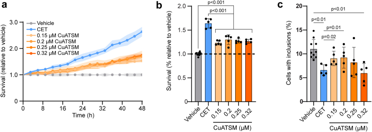
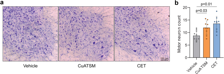
Findings: CET polytherapy reduced inclusion formation and increased cell survival of NSC-34 cells overexpressing SOD1G93A compared to higher concentrations of CuATSM monotherapy. In addition, CET administration was bioavailable and tolerable in mice. CET treatment in SOD1G93A mice delayed disease onset, reduced motor impairments, and increased survival compared to vehicle- and CuATSM-treated mice. In line with these findings, biochemical analysis of lumbar spinal cords showed CET administration improved SOD1 folding, decreased misfolded SOD1 accumulation, and reduced motor neuron loss.

Interpretation: These findings support CET polytherapy as an advantageous alternative compared to CuATSM monotherapy and highlight the potential of utilising small molecules targeting SOD1 as a polytherapy avenue for the treatment of SOD1-associated ALS.

Funding: This work was supported by a FightMND Drug Development Grant, an Australian National Health and Medical Research Council (NHMRC) Investigator Grant (No. 1194872) and a Motor Neuron Disease Research Institute of Australia Bill Gole Postdoctoral Fellowship.

Keywords: Amyotrophic lateral sclerosis; CuATSM; Ebselen; Motor neuron disease; Polytherapy; SOD1; Telbivudine.


Conflict of interest statement

Declaration of interests P.S.D is named as inventor on intellectual property that relates to this research has been licenced from the University of Melbourne to Collaborative Medicinal Development. Collaborative Medicinal Development licenced intellectual property pertaining to CuATSM from the University of Melbourne where PJC is an employee but not a beneficiary of the licence agreement. P.J.C is an unpaid consultant for Collaborative Medicinal Development LLC. J.S.L received grant funding from Molecular Horizons and the University of Wollongong in the form of a Collaboration Grant (M2024). M.L.B, N.E.F, R.B, A.D, F.D, C.G.C, J.G, L.E.M, L.M and J.J.Y declare no conflicts of interest.

Figures

Fig. 1
Fig. 1
Establishing the effect of CET combination therapy on cell survival of NSC-34 cells expressing SOD1G93A-EGFP. (a) Heatmaps of the 3D checkerboard treatment of NSC-34 cells expressing SOD1G93A-EGFP. The colour scale indicates the degree to which treatment improves survival, and numbers indicate the fold increase in survival relative to the vehicle control. (b) The CET ratios that resulted in the greatest improvements in SOD1G93A-EGFP cell survival (≥2.7 fold) from (a) were compared against CuATSM treatment alone. (c) A ratio of 1:80:250 for the CET combination therapy was selected to move forward for further testing using a range of CuATSM concentrations in NSC-34 cells expressing SOD1G93A-EGFP. The CET combination therapy with a CuATSM concentration of 0.15 μM and 0.2 μM improved the survival of SOD1G93A-expressing cells compared to the equivalent concentration of CuATSM alone (determined by a repeated one-way ANOVA with Tukey’s multiple comparisons post-test. All data represent mean ± SD (n = 3 individual experiments).
Fig. 2
Fig. 2
CET combination therapy is more effective than CuATSM treatment in increasing the survival of cells expressing SOD1G93A and reducing SOD1G93A inclusion formation. (a) NSC-34 cells expressing SOD1G93A-EGFP were treated with either CuATSM (0.15 μM), ebselen (12 μM) and telbivudine (37.5 μM) (CET) combination therapy or CuATSM alone and imaged every 3 h for 48 h on an IncuCyte S3 automated fluorescent microscope and GFP cell counts quantified. (b) The survival of SOD1G93A-EGFP expressing cells was determined by measuring the area under the GFP count curves (a). Solid lines represent mean values with shaded areas representing ± SD (n ≥ 5 technical replicates from at least 3 separate experiments) One-way ANOVA with a Tukey’s multiple comparisons post-test was used to compare statistical significance between treatments. (c) The number of cells containing SOD1G93A-EGFP inclusions was determined by semi-automated analysis of the 48 h time point images from the IncuCyte survival assay. One-way ANOVA with a Tukey’s multiple comparisons post-test was used to compare statistical significance between treatments.
Fig. 3
Fig. 3
CET polytherapy improves SOD1G93A folding but does not alter SOD1 activity in NSC-34 cells. (a) The proportion of disulfide-bonded SOD1 was determined by SDS-PAGE migration of SOD1G93A-EGFP lysates treated with either CET or CuATSM under reducing conditions (+β-merc) and nonreducing conditions (−β-merc). Arrows indicate the position of disulfide-bonded SOD1 (SS) and reduced SOD1 (SH) on the immunoblot. (b) Quantification of the immunoblots from (a) by densitometry. Data shown are mean ± SD (n = 3) and statistical significance was determined by one-way ANOVA with a Tukey’s multiple comparisons post-test. (c) Native-PAGE (top) and in-gel zymography (bottom) of SOD1G93A-tdTomato cell lysates treated with either CET or CuATSM. The position of SOD1 dimers, monomers and endogenous mouse SOD1 are indicated with arrows. (d) Quantification of the tdTomato signal from native-PAGE to determine the proportion of SOD1 dimer present following CET or CuATSM treatment. Data shown are mean ± SD (n = 3) and statistical significance was determined by one-way ANOVA with a Tukey’s multiple comparisons post-test. Asterisks indicate significant difference to the vehicle control. (e) SOD1 activity (normalised to SOD1 protein levels) was measured in cells treated with either CET or CuATSM by quantifying the achromatic bands corresponding to the SOD1G93A dimer from in-gel zymography and normalising to the corresponding tdTomato fluorescence signal from the native-PAGE gel. Data represent mean ± SD (n = 3). One-way ANOVA was used to determine significant differences in SOD1 activity between treatments.
Fig. 4
Fig. 4
CET polytherapy delays disease onset, modifies neurological scoring and extends survival in SOD1G93A mice. The effect of daily oral gavage administration of either vehicle, CuATSM or CET on (a) body weight, (b) age of peak body weight, (c) neurological score, (d) age of disease onset (defined as attaining a neurological score of 1), (e) latency to fall during rotarod task, (f) Kaplain–Meier plot and (g) survival (a, c and e) Solid lines represent mean values with shaded areas representing ± SEM (n = 21–24 per treatment group). (b and d) Data is shown as mean ± SD (n = 20–24 per treatment group). (g) Data is shown as median ± IQR.
Fig. 5
Fig. 5
CET treatments increases in vivo SOD1 maturation and reduces misfolded SOD1 accumulation in SOD1G93A mice. (a and b) The relative levels of SOD1 protein and (c) the proportion of disulfide SOD1 (reduced = SH, oxidised intact = SS) in lumbar spinal cord homogenates of vehicle-, CuATSM- and CET-treated mice as determined by non-reducing immunoblot. (d) SOD1 in-gel zymography and (e) quantification of SOD1 activity normalised to soluble SOD1 levels in lumbar spinal cord homogenates of vehicle-, CuATSM- and CET-treated mice. (f) C4F6 dot blots of lumbar spinal cord homogenates and (g) relative misfolded SOD1 (C4F6) levels in lumbar spinal cord homogenates of vehicle-, CuATSM- and CET-treated mice. Data shown are mean ± SD (n = 11–12/treatment). Data are from 3 technical replicates. One-way ANOVA was used to compare relative differences between vehicle-, CuATSM- and CET-treated SOD1G93A mice.
Fig. 6
Fig. 6
CuATSM and CET treatment provides neuroprotection. (a) Representative micrographs of thionin acetate-stained ventral horn lumbar spinal cord sections and (b) motor neuron counts from vehicle-, CuATSM- and CET-treated SOD1G93A mice. Data shown are mean ± SD (n = 9–10/treatment). One-way ANOVA was used to compare relative differences between vehicle-, CuATSM- and CET-treated SOD1G93A mice.

References

    1. Alsultan A.A., Waller R., Heath P.R., Kirby J. The genetics of amyotrophic lateral sclerosis: current insights. Degener Neurol Neuromuscul Dis. 2016;6:49–64. - PMC - PubMed
    1. Abel O., Powell J.F., Andersen P.M., Al-Chalabi A. ALSoD: a user-friendly online bioinformatics tool for amyotrophic lateral sclerosis genetics. Hum Mutat. 2012;33(9):1345–1351. - PubMed
    1. Wang J., Farr G.W., Hall D.H., et al. An ALS-linked mutant SOD1 produces a locomotor defect associated with aggregation and synaptic dysfunction when expressed in neurons of Caenorhabditis elegans. PLoS Genet. 2009;5(1) - PMC - PubMed
    1. Banci L., Barbieri L., Bertini I., Cantini F., Luchinat E. In-cell NMR in E. coli to monitor maturation steps of hSOD1. PLoS One. 2011;6(8) - PMC - PubMed
    1. Casareno R.L., Waggoner D., Gitlin J.D. The copper chaperone CCS directly interacts with copper/zinc superoxide dismutase. J Biol Chem. 1998;273(37):23625–23628. - PubMed

MeSH terms