. 2025 Jun;301(6):108508.
doi: 10.1016/j.jbc.2025.108508. Epub 2025 Apr 11.

Exploiting FcRn engagement of an albumin-CpG oligonucleotide covalent conjugate for potent TLR9 immune induction

Affiliations

Exploiting FcRn engagement of an albumin-CpG oligonucleotide covalent conjugate for potent TLR9 immune induction

Diego Pilati et al. J Biol Chem. 2025 Jun.

Abstract

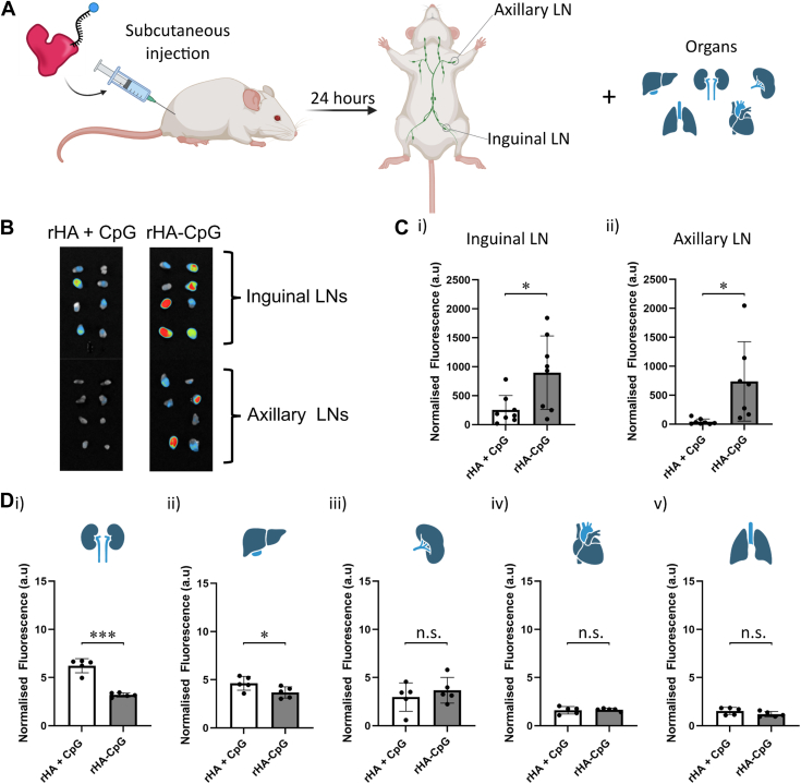
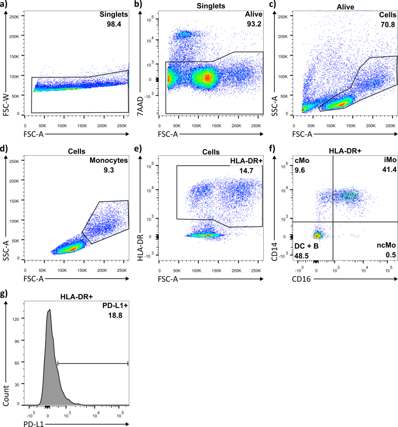
CpG-oligodeoxynucleotide (CpG ODN)-based Toll-like receptor (TLR) agonists are promising immunostimulatory adjuvants; however, low entry into TLR-rich cellular endosomal compartments and poor lymphatic accumulation limit clinical translation. In this work, we introduce a recombinant human serum albumin (rHA)-CpG ODN covalent conjugate (rHA-CpG) designed to exploit the neonatal Fc receptor (FcRn)-driven albumin cellular sorting pathway to maximize CpG delivery into TLR9-rich endosomes and accumulate in lymph nodes. Site-selective conjugation of CpG to albumin cysteine 34, distant from its main FcRn-binding interface, resulted in a retained pH-dependent human FcRn binding, and receptor-driven endosomal trafficking in a cellular recycling assay. Induction of tumor necrosis factor (TNF) secretion in THP-1 cells and interferon alpha (IFN-α) in human hematopoietic stem and progenitor cell (HSPC)-derived plasmacytoid dendritic cells (pDCs), in contrast, to a myeloid differentiation primary response 88 (MyD88) and TLR9 knockout cells, respectively, support TLR9-engagement. The rHA-CpG construct induced greater TNF-α than free CpG ODN in mouse RAW 264.7 cells, and in human peripheral blood mononuclear cells (PBMCs) and expansion of classical (CD14+CD16-) monocytes. Furthermore, greater accumulation of Cy5.5-labelled CpG in the inguinal (>3-fold) and axillary (>18-fold) lymph nodes was observed when conjugated to rHA compared to an unconjugated rHA/CpG mix following subcutaneous injection in mice. Moreover, increased LN accumulation of an rHA variant engineered with high FcRn-binding affinity supports an FcRn-driven mechanism. Demonstration of FcRn-mediated albumin targeting at intra- and extracellular sites provides the mechanistic basis for the potent immune induction observed using the novel rHA-CpG conjugate design class introduced in this work.

Keywords: CpG ODN; FcRn; TLR9; albumin; immune adjuvant; innate immunity; lymph nodes.


Conflict of interest statement

Conflict of interest The authors declare the following financial interests/personal relationships which may be considered as potential competing interests: Kenneth A Howard and Diego Pilati are co-inventors of a patent application on technology described in the paper.

Figures

Figure 1
Figure 1
Proposed mechanism of TLR9 stimulation and FcRn-mediated cellular recycling of rHA-CpG. 1) rHA-CpG uptake in the immune cell endosome. 2) the acidification of the endosome results in albumin-FcRn binding. In the endosomal compartment, the CpG ODN conjugated to rHA interacts with TLR9. 3) TLR9 activation induces the secretion of IFNs and proinflammatory cytokines. 4) rHA-CpG is recycled by FcRn to the cellular membrane through an exocytotic process and is released in the extracellular space upon exposure to physiological pH, thereby evading lysosomal degradation.
Figure 2
Figure 2
PURITY and FcRn affinity of rHA-CpG.A, schematic illustration showing conjugation of (i) SM(PEG)8 to amine-modified CpG ODN, and (ii) maleimide-modified CpG ODN to rHA. B, native-PAGE of CpG ODN (1), CpG ODN + linker (2), rHA (3), and rHA-CpG (4) stained with SYBR Gold and Coomassie Blue. (n = 1). C, Size Exclusion Chromatography (SEC) of purified rHA and rHA-CpG. (n = 1). D, binding affinity of rHA WT, rHA HB, rHA-CpG WT, and rHA-CpG HB to FcRn at pH 5.5 measured by ELISA. (n = 3). E, detection of recombinant albumin in supernatant from human FcRn expressing cells incubated with rHA and rHA-CpG (n = 3). n = represents the number of independent repeats for each experiment. Statistical analysis was made using an unpaired t test, ∗∗∗∗p < 0.0001. Error bars represent standard deviation.
Figure 3
Figure 3
Investigation of rHA-cpG mechanism and cell immune stimulation.A, schematic illustration showing mechanism of TLR9 signaling in THP-1 WT and THP-1 MyD88 KO cells. B, fold change in the expression of TNF-α induced with 0.25 μM of either CpG (Class C) ODN, rHA-ODN, rHA-CpG, rHA, and rHA + CpG in THP-1 WT and (C) THP-1 MyD88 KO (n = 3). D, secretion of TNF-α induced with 0.25 μM of either ODN, CpG ODN, rHA-ODN, and rHA-CpG from murine RAW 264.7 cells. (n = 3 for untreated, ODN and rHA-ODN, n = 4 for CpG ODN and rHA-CpG). E, secretion of TNF-α induced with 10 nM of either CpG ODN or rHA-CpG, or with 0.5 μg/ml LPS, from human PBMCs. (n = 3). n = represents the number of independent repeats for each experiment. For all experiments, the statistical analysis was made using a one-way ANOVA with multiple comparisons and Tukey's post hoc correction, ∗p < 0.05, ∗∗∗p < 0.001, ∗∗∗∗p < 0.0001, n.s. = not significant. Error bars represent standard deviation.
Figure 4
Figure 4
Effect of rHA-CpG on proliferation and differentiation of PBMCs. PBMCs were exposed to 10 nM CpG (Class C) ODN, 10 nM rHA-CpG, or to 0.5 μg/ml LPS. RPMI was used for cell only control. A, percentage of the monocyte population in PBMCs at 24, 48 and 72 h, normalized to the cells only control at 24 h. (n = 3). B, percentage of HLA-DR expressing cells in PBMCs at 24, 48 and 72 h, normalised to the cells only control at 24 h. C, percentage of DC + B cells and monocyte subsets cMo, iMo and ncMo in HLA-DR+ cells at 24, 48 and 72 h. D, line graph representation of the normalized percentage of cMo cells at 24, 48 and 72 h from Figure 3D. n = represents the number of independent repeats for each experiment. For all experiments, statistical analysis was made using a two-way ANOVA with multiple comparisons and Tukey's post hoc correction, ∗p < 0.05, ∗∗p < 0.01, ∗∗∗∗p < 0.0001. Light blue asterisk represent significance between rHA-CpG and CpG ODN. Error bars represent standard deviation.
Figure 5
Figure 5
Lymph node accumulation and biodistribution of rHA-CpG.A, schematic representation of the in vivo protocol. C57BL/6 mice were injected subcutaneously at the tail base with rHA-CpG, rHA + CpG or PBS (n = 5 per group) with each group receiving 1.2 nmol of CpG (Class B ODN labeled with Cy5.5). After 24 h, draining (inguinal) and distal (axillary) LNs were collected for imaging. Fluorescence values for lymph nodes were normalized to the PBS control. B, Fluorescence images of excised draining (inguinal) and distal (axillary) LNs from 4 animals per group (n = 8 LNs in total [a pair taken from each animal]). Fluorescence quantification of the Cy5.5 labelled CpG in Ci) Inguinal (draining) LNs Cii) Axillary (distal) LNs, measurements normalised to the PBS control. Fluorescence quantification of the Cy5.5 labelled CpG in Di) kidneys Dii) liver Diii) spleen Div) heart Dv) lungs, measurement normalised to the PBS control and organ weight. The statistical analysis was made using an unpaired t test, ∗p < 0.05, ∗∗p < 0.01, n.s. = not significant. Error bars represent standard deviation. Created with Biorender.com.
Extended data Figure 1
Extended data Figure 2
Extended data Figure 3
Extended data Figure 4
Extended data Figure 5

References

    1. Li D., Wu M. Pattern recognition receptors in health and diseases. Signal. Transduct. Target Ther. 2021;6:291. - PMC - PubMed
    1. Kawasaki T., Kawai T. Toll-like receptor signaling pathways. Front. Immunol. 2014;5:461. - PMC - PubMed
    1. Fitzgerald K.A., Kagan J.C. Toll-like receptors and the control of immunity. Cell. 2020;180:1044–1066. - PMC - PubMed
    1. Hornung V., Rothenfusser S., Britsch S., Krug A., Jahrsdörfer B., Giese T., et al. Quantitative expression of toll-like receptor 1-10 mRNA in cellular subsets of human peripheral blood mononuclear cells and sensitivity to CpG oligodeoxynucleotides. J. Immunol. 2002;168:4531–4537. - PubMed
    1. Kadowaki N., Ho S., Antonenko S., Malefyt R.W., Kastelein R.A., Bazan F., et al. Subsets of human dendritic cell precursors express different toll-like receptors and respond to different microbial antigens. J. Exp. Med. 2001;194:863–869. - PMC - PubMed

MeSH terms

LinkOut - more resources