. 2025 Apr 14;15(1):12755.
doi: 10.1038/s41598-025-97584-1.

Leukoreduction filter derived NK cells offer a promising source for off the shelf CAR NK cell immunotherapy

Affiliations

Leukoreduction filter derived NK cells offer a promising source for off the shelf CAR NK cell immunotherapy

Ali Moazzeni et al. Sci Rep. .

Abstract

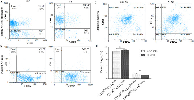
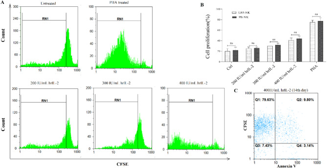
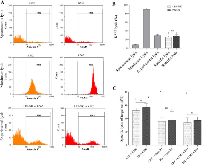
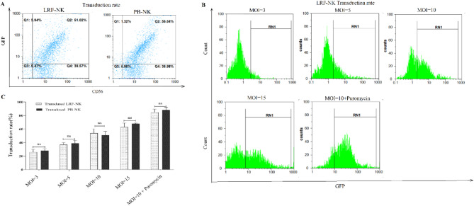
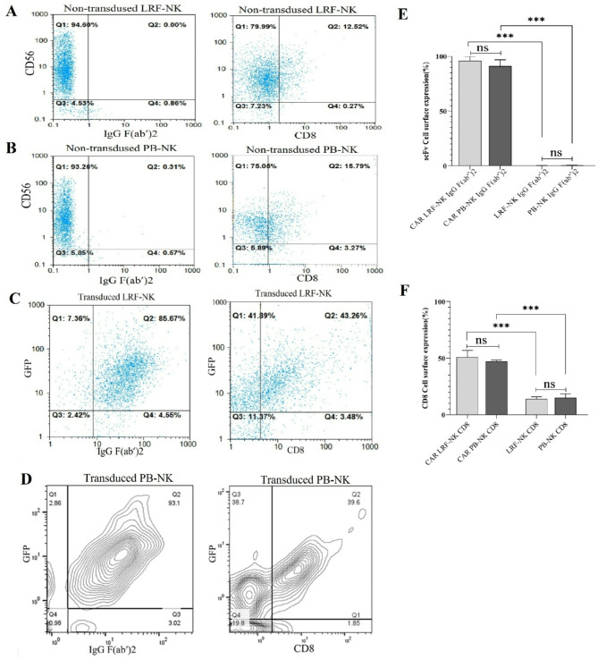
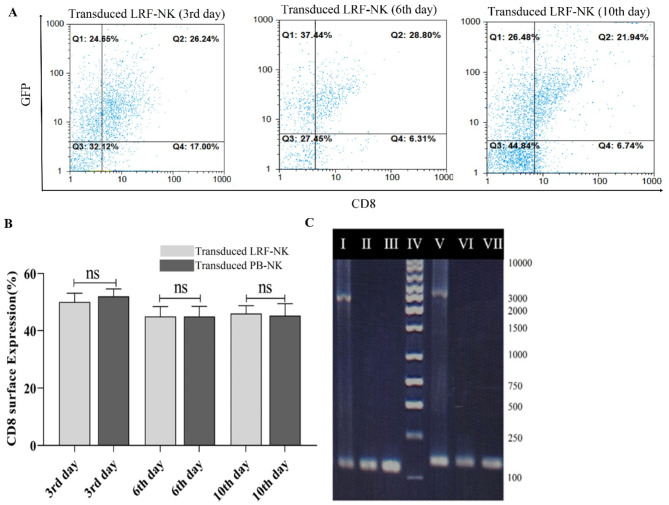
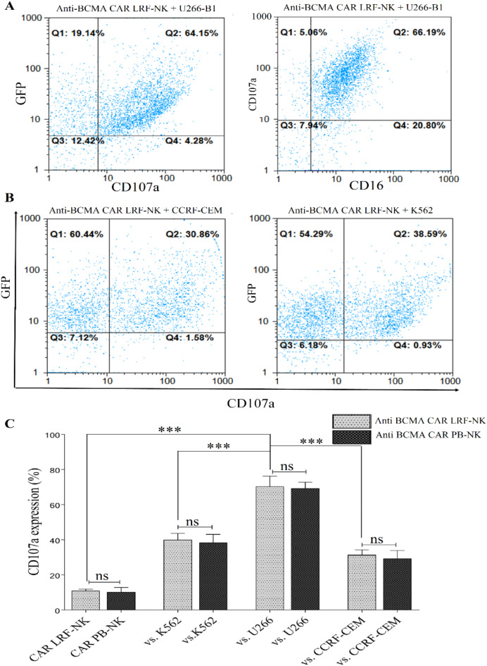
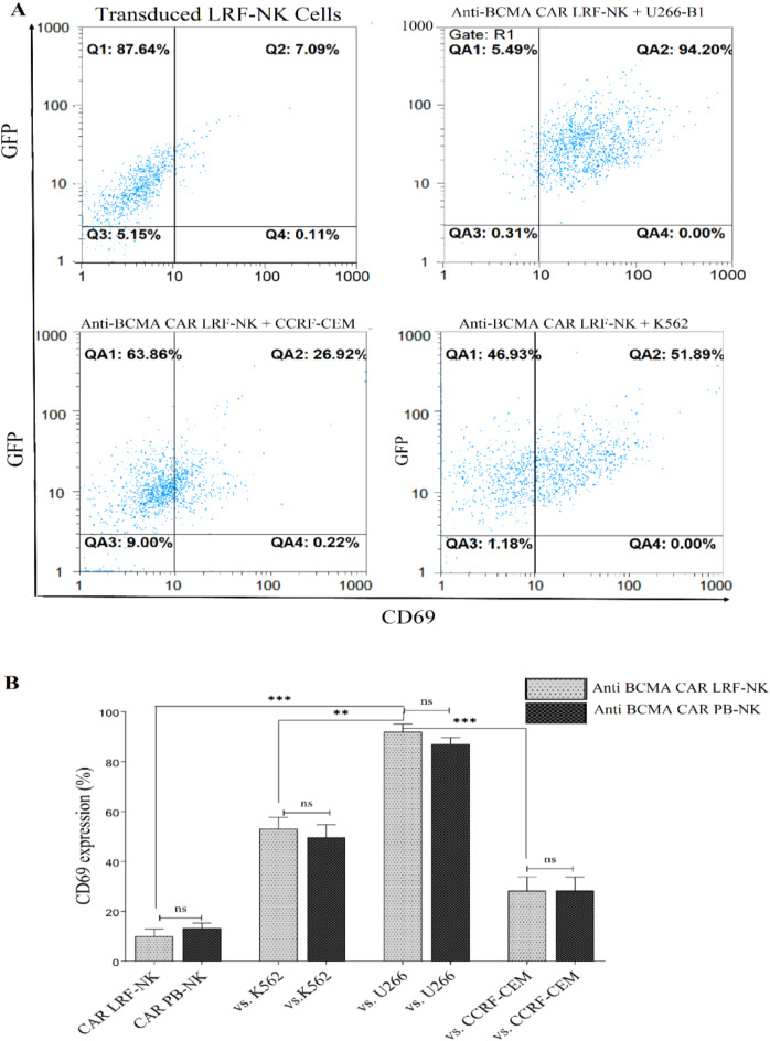
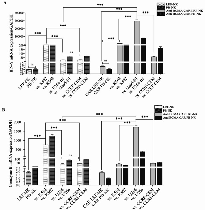
Immunotherapy employing natural killer (NK) cells has emerged as a transformative approach to treating hematological malignancies. The reprogramming of NK cells by incorporating a chimeric antigen receptor (CAR) equipped with potent signaling domains has demonstrated efficacy in enhancing NK cell responses and improving specificity in recognizing cancerous cells. Despite these advancements, the primary challenge in implementing allogeneic NK cell therapy requiring a viable donor source for clinically relevant doses remains unresolved. This study tested NK cells obtained from leukoreduction filters (LRF) post-blood donation to address the need for an efficient and scalable supply of NK cells for generating anti-BCMA CAR NK cells. LRF-NK cells were isolated under sterile conditions and compared with peripheral blood (PB)-derived NK cells in terms of immunophenotype, proliferation capacity, and functional characteristics. Notably, no significant differences in inherent characteristics were observed between LRF-NK and PB-NK cells. Subsequently, both NK cell populations were employed to generate anti-BCMA CAR-NK cells. The data revealed a high specific cytotoxicity of Anti-BCMA CAR LRF-NK cells during co-culture with U266-B1 cells (70.3 ± 4.78%), surpassing that observed with CCRF-CEM cells (31.3 ± 2.35%) and similar to Anti-BCMA CAR PB-NK cells. Furthermore, the expression of IFN-γ and Granzyme B, following the co-culture of Anti-BCMA CAR LRF-NK cells with target cells, mirrored that observed in Anti-BCMA CAR PB-NK cells. This study provides the rationale and feasibility of utilizing LRF-NK cells as a safe, high-yield, accessible, and optimal cost-effective source for cancer immunotherapy.

Keywords: Chimeric antigen receptor (CAR); Healthy donor; Leukoreduction filters (LRF); Natural killer (NK) cell.


Conflict of interest statement

Declarations. Competing interests: The authors declare no competing interests.

Figures

Fig. 1
Fig. 1
The purity and phenotype of LRF-NK and PB-NK cells. (A) Pre-MACS NK cell isolation: phenotypic analysis of anti-CD3 and anti-CD56-labeled isolated lymphocytes from LRF and PB. (B) Post-MACS separation: Purified LRF-NK and PB-NK cells characterized based on CD56 expression and the absence of CD3 expression. (C) CD16 and CD56 phenotypic expression on LRF-NK and PB-NK cells post-MACS separation. (D) Statistical comparison reveals no significant differences in the immunophenotype of NK cells from LRF-NK and PB-NK, considering populations with CD56dimCD16bright and CD56brightCD16dim/neg phenotypes (P = 0.14 and P = 0.32, respectively). LRF: leukocyte reduction Filter, PB: peripheral blood.
Fig. 2
Fig. 2
Proliferation and Viability Comparison of LRF and PB-NK Cells. (A) The dose-dependent proliferation of LRF-NK cells with 200, 300, and 400 IU/mL hrIL-2 treatment on day 14 was detected by CFSE staining. Untreated and PHA-treated LRF-NK cells served as negative and positive controls, respectively. LRF-NK cells, undergoing up to two, three and four division cycles following induction with 200, 300 and 400 IU doses of IL-2 respectively. (B) Statistical analysis reveals no significant differences in proliferation capacity between LRF-NK and PB-NK cells treated with different hrIL-2 concentrations. (C) The LRF-NK cell viability following treatment with 400 IU/mL IL-2 was detected using the annexin V on day 14. Data presented as mean ± SD. ** Represents P < 0.001 and *** P < 0.0001 (N = 3). LRF: Leukocyte reduction filter, PB: peripheral blood, PHA: phytohemagglutinin, CFSE: carboxyfluorescein diacetate succinimidyl ester.
Fig. 3
Fig. 3
The cytotoxic effect of LRF-NK and PB-NK cells against CFSE-labeled target cell lines. (A) Flow cytometric representation of spontaneous and maximum lysis of the K562 cell line and experimental lysis following co-culture with LRF-NK cells was detected by Annexin V and 7AAD staining. (B) Statistical comparison of spontaneous, maximum, experimental, and specific lysis of the K562 cell line. (C) Mean percentages of specific lysis for K562, U266-B1, and CCRF-CEM cell lines following co-culture with LRF-NK and PB-NK cells. A significant increase in K562-specific lysis was observed compared to U266-B1 (P = 0.0184) and CCRF-CEM cells (P = 0.0194). No significant difference was observed in the specific lysis of U266-B1 and CCRF-CEM cell lines (P = 0.7306). LRF-NK and PB-NK cells showed no significant differences in inducing specific lysis across the three mentioned cell lines (P > 0.05). Data are presented as mean ± SD. (** rEpresents P < 0.001 and *** P < 0.0001, N = 3). LRF: Leukocyte reduction filter, PB: peripheral blood.
Fig. 4
Fig. 4
Transduction rate of LRF-NK and PB-NK cells. (A) Flow cytometry representation of the transduction rate of LRF-NK and PB-NK cells, detected through GFP expression. (B) Flow cytometric visualization of the transduction rate of LRF-NK cells at MOIs of 3, 5, 10, 15, and 10 supplemented with puromycin. (C) Statistical analysis revealed an augmented transduction rate with increasing MOI and puromycin treatment. No significant differences were observed in the transduction rates between LRF-NK and PB-NK cells (P > 0.05). Data are presented as mean ± SD. (** represents P < 0.001 and *** P < 0.0001, N = 3). LRF: leukocyte reduction filter, pb: peripheral blood.
Fig. 5
Fig. 5
Anti-BCMA CAR expression on LRF-NK and PB-NK cells, emphasizing key aspects of scFv and CD8 expression. (A) Non-transduced LRF-NK cells lack scFv expression and low CD8 surface expression, detected by anti-IgG (Fab’)2 and Anti CD8 antibodies. (B) Similarly, non-transduced PB-NK cells show no scFv expression, detected by anti-IgG (Fab’)2 and low CD8 surface expression. (C) High expression of scFv and CD8 on GFP + transduced LRF-NK cells. (D) The scFv expressions by GFP+ transduced LRF NK cells are illustrated through the contour plot demonstration a significantly increased expression. (E) The scFv expressions by non-transduced LRF/PB NK cells in comparison with GFP+ transduced LRF/PB NK cells are illustrated through the contour plot demonstration a significantly increased expression (F). The CD8 expressions by non-transduced LRF/PB NK cells in comparison with GFP+ transduced LRF/PB NK cells are illustrated through the contour plot demonstration a significantly increased expression. LRF: leukoreduction filters, PB: peripheral blood.
Fig. 6
Fig. 6
Persistent Expression of Anti-BCMA CAR in LRF-NK Cells and Integration of CAR Construct into NK Cells. (A) Assessment of the sustained expression of the CAR construct through detecting the CD8 marker on GFP + transduced LRF-NK cells at days 3, 6, and 10 post-transductions. (B) Mean percentage of CD8 expression on transduced LRF-NK cells compared to PB-NK cells on days 3, 6, and 10 post-transduction. (C) Electrophoresis plot illustrating the integration of the CAR gene into NK cells. I: CD3ζ positive control, II: CD3ζ (CAR LRF-NK), III: CD3ζ (PB-NK), IV: 100 bp Ladder, V: scFv positive control, VI: scFv (CAR LRF-NK), VIII: scFv (CAR PB-NK).
Fig. 7
Fig. 7
High cytotoxicity of anti-BCMA CAR LRF-NK and PB-NK cells against BCMA-expressing cells. (A) Flow cytometric analysis depicts CD107a expression on anti-BCMA CAR LRF-NK cells (GFP+ population) post-co-culture with U266-B1 cells (BCMA+ cells). (B) the mean percentage of CD107a expression on anti-BCMA CAR LRF-NK cells (GFP+ population) after co-culture with CCRF-CEM (BCMA cells) and K562 cell lines. (C) Statistical analysis indicates significantly elevated CD107a expression on anti-BCMA CAR LRF- and PB-NK cells when co-cultured with U266-B1 compared to CCRF-CEM and K562 cells. No significant difference in CD107a expression exists between anti-BCMA CAR LRF- and PB-NK cells. Data presented as Mean ± SD. (** Represents P < 0.001, and *** P < 0.0001, N = 3). LRF: Leukocyte reduction filter. PB: peripheral blood.
Fig. 8
Fig. 8
Specific activation of transduced NK cells. (A) The baseline expression levels of CD69 (upper left) on anti-BCMA CAR LRF-NK cells, when cultured alone and CD69 expression following the co-cultivation of anti-BCMA CAR LRF-NK cells with U266-B1 (upper right) CCRF-CEM (Lower Left) and K562 (lower left) cell lines. (B) The mean percentage of CD69 as an early activation marker significantly increased on anti-BCMA CAR LRF/PB cells following co-cultivation with the U266-B1 compared to CCRF-CEM cell lines.
Fig. 9
Fig. 9
mRNA expression of IFN-γ (A) and granzyme B (B) in anti-BCMA CAR LRF-NK and PB-NK cells following co-culture with U266-B1 (BCMA+), CCRF-CEM (BCMA), and K562 Cell Lines. The augmented expression of IFN-γ and granzyme B was evident in LRF-NK and PB-NK cells after co-cultivation with the K562 cell line, as opposed to their counterparts co-cultivated with U266-B1 and CCRF-CEM cell lines, as well as IL-2 treated NK cells. No statistically significant distinctions were discerned between LRF-NK and PB-NK cells. Conversely, Anti-BCMA CAR NK cells demonstrated a notable increase in the expression of IFN-γ and GrB after co-culture with U266-B1 compared to CCRF-CEM. The absence of significant differences between LRF and PB CAR NK cells in IFN-γ and GrB expression under identical conditions is noteworthy. Notably, a significantly heightened expression of IFN-γ and GrB in CAR NK cells compared to regular NK cells was observed after co-culture with U266-B1 cells. Data are presented as mean ± SD. (Double asterisks (**) signify P < 0.001, and triple asterisks (***) denote P < 0.0001; N = 3). LRF: leukocyte reduction filter. PB: peripheral blood.

Similar articles

References

    1. Shin, M. H. et al. NK cell-based immunotherapies in cancer. Immune Netw.20(2) (2020). - PMC - PubMed
    1. Feins, S., Kong, W., Williams, E. F., Milone, M. C. & Fraietta, J. A. An introduction to chimeric antigen receptor (CAR) T-cell immunotherapy for human cancer. Am. J. Hematol.94(S1), S3–S9 (2019). - PubMed
    1. Xie, G. et al. CAR-NK cells: A promising cellular immunotherapy for cancer. EBioMedicine59, 102975 (2020). - PMC - PubMed
    1. Miliotou, A. N. & Papadopoulou, L. C. CAR T-cell therapy: a new era in cancer immunotherapy. Curr. Pharm. Biotechnol.19(1), 5–18 (2018). - PubMed
    1. Hu, Y., Tian, Z. & Zhang, C. Chimeric antigen receptor (CAR)-transduced natural killer cells in tumor immunotherapy. Acta Pharmacol. Sin.39(2), 167 (2018). - PMC - PubMed

MeSH terms

LinkOut - more resources