. 2025 Apr 13;24(1):24.
doi: 10.1186/s12941-025-00790-y.

Novel strategies for vancomycin-resistant Enterococcus faecalis biofilm control: bacteriophage (vB_EfaS_ZC1), propolis, and their combined effects in an ex vivo endodontic model

Affiliations

Novel strategies for vancomycin-resistant Enterococcus faecalis biofilm control: bacteriophage (vB_EfaS_ZC1), propolis, and their combined effects in an ex vivo endodontic model

Toka A Hakim et al. Ann Clin Microbiol Antimicrob. .

Abstract

Background: Endodontic treatment failures are predominantly attributed to Enterococcus faecalis (E. faecalis) infection, a Gram-positive coccus. E. faecalis forms biofilms, resist multiple antibiotics, and can withstand endodontic disinfection protocols. Vancomycin-resistant strains, in particular, are challenging to treat and are associated with serious medical complications.

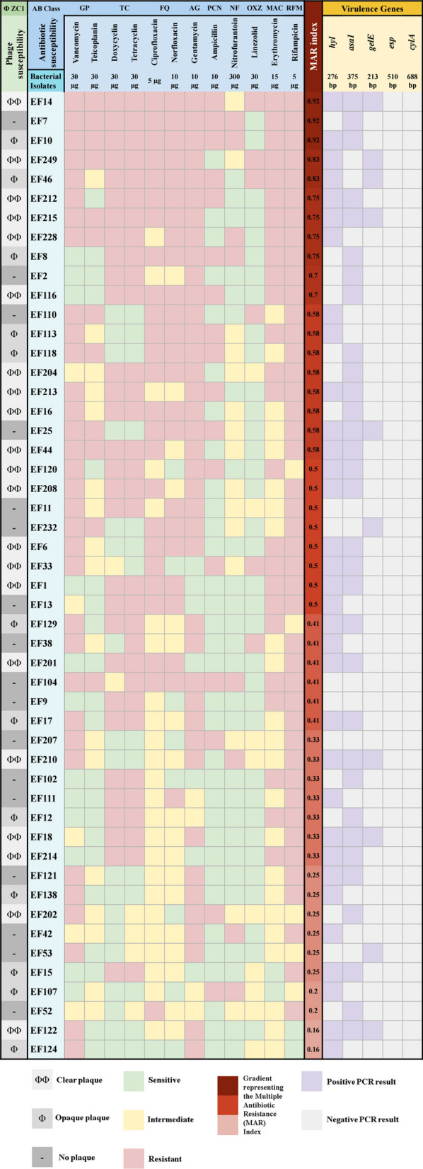
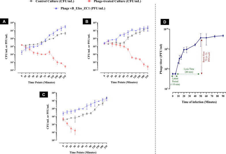
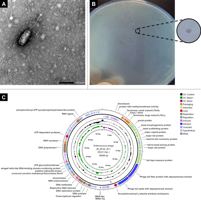
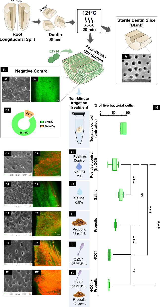
Methods: A novel phage, vB_EfaS_ZC1, was isolated and characterized. Its lytic activity against E. faecalis was assessed in vitro through time-killing and biofilm assays. The phage's stability under various conditions was determined. Genomic analysis was conducted to characterize the phage and its virulence. The phage, propolis, and their combination were evaluated as an intracanal irrigation solution against a 4-week E. faecalis mature biofilm, using an ex vivo infected human dentin model. The antibiofilm activity was analyzed using a colony-forming unit assay, field emission scanning electron microscopy, and confocal laser scanning microscopy.

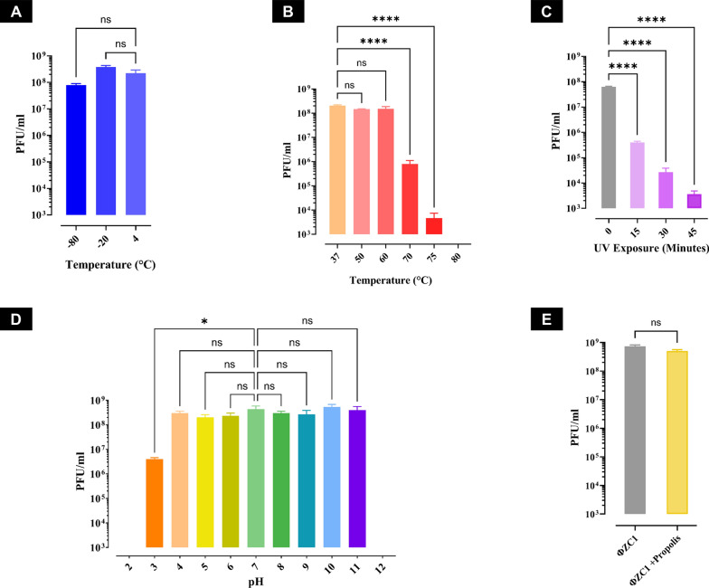
Results: The isolated phage, vB_EfaS_ZC1, a siphovirus with prolate capsid, exhibited strong lytic activity against Vancomycin-resistant strains. In vitro assays indicated its effectiveness in inhibiting planktonic growth and disrupting mature biofilms. The phage remained stable under wide range of temperatures (- 80 to 60 °C), tolerated pH levels from 4 to 11; however the phage viability significantly reduced after UV exposure. Genomic analysis strongly suggests the phage's virulence and suitability for therapeutic applications; neither lysogeny markers nor antibiotic resistance markers were identified. Phylogenetic analysis clustered vB_EfaS_ZC1 within the genus Saphexavirus. The phage, both alone and in combination with propolis, demonstrated potent antibiofilm effects compared to conventional root canal irrigation.

Conclusion: Phage vB_EfaS_ZC1 demonstrates a promising therapy, either individually or in combination with propolis, for addressing challenging endodontic infections caused by E. faecalis.

Keywords: Saphexavirus; Antibiofilm; Endodontic treatment; Irrigation; Phage therapy; Propolis; Vancomycin-resistant Enterococcus faecalis.


Conflict of interest statement

Declarations. Ethics approval and consent to participate: The Preferred Reporting Items for Laboratory Studies in Endodontology (PRILE) 2021 standards were followed in writing the paper for this laboratory study. This study was approved by the Research Ethics Committee (REC), Number 703/2023. Faculty of Dentistry, Suez Canal University, Ismailia, Egypt. Established according to “WHO-2011” standards (Date of approval: 3 October 2023). Consent for publication: Not applicable. Competing interests: The authors declare that they have no competing interests.

Figures

Fig. 1
Fig. 1
Heatmap illustrating the susceptibility to phage vB_EfaS_ZC1 and antimicrobial agents, and virulence gene profiles of 50 bacterial isolates. The left panel describes the host range of phage vB_EfaS_ZC1 visualized by plaque morphology: clear lysis (ΦΦ), opaque lysis (Φ), and no lysis (-) for each bacterial isolate. The middle panel represents the antimicrobial susceptibility profiles using color coding: green for sensitive, yellow for intermediate, and red for resistant isolates. A gradient heatmap in shades of red represents the Multiple Antibiotic Resistance (MAR) Index, with darker shades indicating higher indices. The right panel presents the PCR detection results for five virulence genes, with purple and light gray indicating positive and negative results, respectively
Fig. 2
Fig. 2
Stability assessment of phage vB_EfaS_ZC1 under various conditions. A Storage temperatures stability at − 80 °C, − 20 °C, and 4 °C. B Thermal stability of phage vB_EfaS_ZC1 at temperatures range from 37 to 80 °C. C Stability of the phage vB_EfaS_ZC1 under UV exposure assessed at an interval of 15 min for 45 min. D Stability of phage vB_EfaS_ZC1 across a pH range of 2–12. E Stability of phage vB_EfaS_ZC1 in the presence and absence of propolis (12 µg/mL), represented after a 3 h incubation period. Error bars represent the standard deviation, and statistical significance are indicated as ns for no significant difference (P ≥ 0.05), *indicates (P < 0.05), and ****indicates (P < 0.0001)
Fig. 3
Fig. 3
In vitro bacteriolytic dynamics of phage vB_EfaS_ZC1 at different MOIs. The three panels (A, B, and C) represent EF/14 cultures infected with phage vB_EfaS_ZC1 at MOIs 0.1, 1, and 10, respectively. These panels illustrate bacterial counts (CFU/mL) and phage titer (PFU/mL) over a period of 210 min. D One-step growth curve of phage vB_EfaS_ZC1 at MOI 0.1
Fig. 4
Fig. 4
Antibiofilm activity of phage vB_EfaS_ZC1 at different MOIs. A Quantification of biofilm inhibition in bacterial cultures treated with phage vB_EfaS_ZC1 at MOIs (0.001–100), measured as OD 595 nm. B Quantification of biofilm clearance in preformed biofilms treated with phage vB_EfaS_ZC1 at MOIs (0.001–100), measured as OD 595 nm. Statistical significance between phage-treated groups and the untreated group is indicated * and ****which represent P < 0.05 and P < 0.0001, respectively
Fig. 5
Fig. 5
Morphological characteristics of phage vB_EFaS_ZC1. A Transmission electron micrograph of phage vB_EFaS_ZC1 captured using TEM B Phage vB_EFaS_ZC1 plaques formed in double-layer agar plates on the EF/14 lawn. C Circle map visualization of phage vB_EfaS_ZC1 genome. Colored segments represent coding sequences categorized by predicted function: orange (genome packaging), light green (assembly of virion proteins), red (lysis), brown (replication), turquoise (regulation), pink (immune), blue (infection), light blue (unsorted/hypothetical), and grey (tRNA gene). The middle circle displays GC content (black). The inner circle shows a GC skew with green for the positive skew and magenta for the negative skew
Fig. 6
Fig. 6
Phylogenetic analysis of phage vB_EfaS_ZC1. Three panels represent the evolutionary relationships of phage vB_EfaS_ZC1: A A proteomic tree with the phage marked by a red star, the circular tree comparing vB_EFaS_ZC1 to all phages in the ViPtree database, and the rectangular tree highlighting closely related phages with high ViPtree similarity scores (SG > 0.6). B VIRIDIC heatmap visualizing the intergenomic similarity of vB_EFaS_ZC1 (red star) to closely related phages, all members of the Saphexavirus genus based on NCBI taxonomy; Saphexaviruses in ICTV marked by blue circles. The heatmap represents clustering at genus and species levels. C A maximum likelihood phylogenetic tree comparing the terminase large subunit (TerL, core protein) of vB_EfaS_ZC1 (red triangle) with homologs from other closely related Saphexavirus phages. Four phages with low SG scores (< 0.6) to vB_EfaS_ZC1 were chosen as an outgroup. Phylogeny.fr was used for tree construction
Fig. 7
Fig. 7
Evaluation of the antibacterial efficacy of phage, propolis, and their combination against EF/14 bacteria in planktonic and biofilm forms. A Time-killing curve of EF/14 culture treated with phage vB_EfaS_ZC1, propolis, and their combination over 48 h. B Efficacy of different irrigation treatments against EF/14 biofilms on dentin slices. The statistical significance of the reduction in bacterial count (CFU/mL) after different irrigation treatments compared to the control (untreated) group is represented by ** for P < 0.01 and **** for P < 0.0001
Fig. 8
Fig. 8
Characterization of EF/14 biofilm morphology and viability following irrigation treatments in ex vivo model of root dentin slices. A Sterilized blank group without biofilm, while (B) contained a four-week-old biofilm without any treatment. (C- G) underwent different irrigated treatments: (C) was treated with 2% Sodium Hypochlorite, (D) with 0.9% saline, (E) with propolis, (F) with phage vB_EfaS_ZC1, and (G) with a combination of phage vB_EfaS_ZC1 and propolis. These groups are represented in the corresponding figure. B1-G1 Qualitative analysis was observed by FESEM images of the EF/14 biofilm after 10 min of different irrigation treatments. B2–G2 Quantitative analysis by CLSM illustrating EF/14 biofilms (green: live bacteria; red: dead bacteria) on dentin slices after 10 min of different irrigation treatments. H Box plots demonstrate the percentage of live bacterial cells under different irrigation treatments. The median and interquartile range (n = 6) are represented for each group, Statistical significance is indicated by *** for P < 0.001, and ns indicates no significant difference.

Similar articles

References

    1. Alghamdi F, Shakir M. The influence of Enterococcus faecalis as a dental root canal pathogen on endodontic treatment: a systematic review. Cureus. 2020;12(3): e7257. - PMC - PubMed
    1. Haapasalo M, et al. Eradication of endodontic infection by instrumentation and irrigation solutions. Endod Top. 2005;10(1):77–102.
    1. van Harten RM, et al. Multidrug-resistant Enterococcal Infections: New compounds, novel antimicrobial therapies? Trends Microbiol. 2017;25(6):467–79. - PubMed
    1. Ali, L., et al., Molecular Mechanism of Quorum-Sensing in Enterococcus faecalis: Its Role in Virulence and Therapeutic Approaches. Int J Mol Sci, 2017. 18(5). - PMC - PubMed
    1. Hashem YA, Abdelrahman KA, Aziz RK. Phenotype-genotype correlations and distribution of key virulence factors in enterococcus faecalis isolated from patients with urinary tract infections. Infect Drug Resist. 2021;14:1713–23. - PMC - PubMed

MeSH terms

LinkOut - more resources