. 2025 Apr 14;24(1):113.
doi: 10.1186/s12943-025-02319-5.

BIRC2 blockade facilitates immunotherapy of hepatocellular carcinoma

Affiliations

BIRC2 blockade facilitates immunotherapy of hepatocellular carcinoma

Lingyi Fu et al. Mol Cancer. .

Abstract

Background: The effectiveness of immunotherapy in hepatocellular carcinoma (HCC) is limited, however, the molecular mechanism remains unclear. In this study, we identified baculoviral IAP repeat-containing protein 2 (BIRC2) as a key regulator involved in immune evasion of HCC.

Methods: Genome-wide CRISPR/Cas9 screening was conducted to identify tumor-intrinsic genes pivotal for immune escape. In vitro and in vivo models demonstrated the role of BIRC2 in protecting HCC cells from immune killing. Then the function and relevant signaling pathways of BIRC2 were explored. The therapeutic efficacy of BIRC2 inhibitor was examined in different in situ and xenograft HCC models.

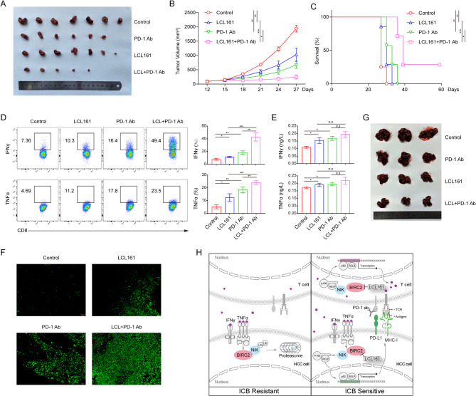
Results: Elevated expression of BIRC2 correlated with adverse prognosis and resistance to immunotherapy in HCC patients. Mechanistically, BIRC2 interacted with and promoted the ubiquitination-dependent degradation of NFκB-inducing kinase (NIK), leading to the inactivation of the non-canonical NFκB signaling pathway. This resulted in the decrease of major histocompatibility complex class I (MHC-I) expression, thereby protecting HCC cells from T cell-mediated cytotoxicity. Silencing BIRC2 using shRNA or inhibiting it with small molecules increased the sensitivity of HCC cells to immune killing. Meanwhile, BIRC2 blockade improved the function of T cells both in vitro and in vivo. Targeting BIRC2 significantly inhibited tumor growth, and enhanced the efficacy of anti-programmed death protein 1 (PD-1) therapy.

Conclusions: Our findings suggested that BIRC2 blockade facilitated immunotherapy of HCC by simultaneously sensitizing tumor cells to immune attack and boosting the anti-tumor immune response of T cells.

Keywords: BIRC2; Hepatocellular carcinoma; Immunotherapy; MHC-I; NIK.


Conflict of interest statement

Declarations. Ethics approval and consent to participate: Ethical approval was granted by the Sun Yat-sen University Cancer Center Institute Research Ethics Committee, and all procedures adhered to the International Ethical Guidelines for Biomedical Research Involving Human Subjects (CIOMS) (GZR2022-054). Written informed consent was obtained from each patient or their legal guardians. Ethical approval for all animal procedures was granted by the Institutional Animal Care and Use Committee of Sun Yat-sen University (L102012022005T). Competing interests: The authors declare no competing interests.

Figures

Fig. 1
Fig. 1
BIRC2 is a tumor-intrinsic immune evasion gene. A. Schematic representation of the CRISPR/Cas9 screening process. B. Volcano plot illustrating the normalized fold change of sgRNA in Hepa1-6-OVA cells challenged with OT-I T cells compared to control CD8+ T cells, with selected top candidates highlighted. C. Schematic representation of the competitive assay. D. Apoptosis of Hepa1-6-OVA cells infected with sgBIRC2 after challenging with OT-I T cells. E. Clone formation of Hepa1-6-OVA cells infected with sgBIRC2 after challenging with OT-I T cells. Representative of three independent experiments. Statistical analysis was performed on biological replicates (n = 3); each value represents mean ± SD; two-sided Student t test. *, P < 0.05; **, P < 0.01; ***, P < 0.001. F. Tumor growth of Hepa1-6-OVA cells in NSG and C57BL/6J mice. Representative of two independent experiments (n = 6–9 mice/group); each value represents mean ± SEM. Survival analysis performed using the log-rank (Mantel-Cox) test. *, P < 0.05; **, P < 0.01; ***, P < 0.001
Fig. 2
Fig. 2
BIRC2 expression correlates with poor survival and resistance to ICB in HCC patients. A. Representative HE and IHC images of BIRC2 in HCC tissues. B. Representative IHC images of CD8 and CD4 in high- and low-BIRC2 groups. C. The number of CD8+ and CD4+ T cells in high- and low-BIRC2 groups. D. Representative images of tumor-infiltrating lymphocytes in high- and low-BIRC2 groups. E. Kaplan-Meier analysis of the OS comparing high and low BIRC2 expression in HCC patients (n = 765). F. Kaplan-Meier analysis of the DFS comparing high and low BIRC2 expression in HCC patients (n = 765). G. Kaplan-Meier analysis of OS comparing high and low BIRC2 expression in HCC patients who received combined immunotherapy (n = 144). H. Kaplan-Meier analysis of PFS comparing high and low BIRC2 expression in HCC patients who were treated with anti-PD-1 plus Lenvatinib (n = 47). Representative of three independent experiments. Statistical analysis was performed on biological replicates (n = 3); each value represents mean ± SD; two-sided Student t test. *, P < 0.05; **, P < 0.01; ***, P < 0.001
Fig. 3
Fig. 3
BIRC2 suppresses the expression of HLA-ABC and PD-L1 induced by IFNγ. A. KEGG analysis of significant pathways up-regulated in PLC cells infected with shBIRC2. B. Expression of HLA-ABC and PD-L1 in PLC cells infected with shBIRC2 and treated with IFNγ. C. Expression of HLA-ABC and PD-L1 in PLC cells infected with shBIRC2 and cultured in conditioned medium. D. Membrane expression of HLA-ABC and PD-L1. E. Expression and subcellular localization of HLA-ABC and PD-L1. Representative of three independent experiments. Statistical analysis was performed on biological replicates (n = 3); each value represents mean ± SD; two-sided Student t test. *, P < 0.05; **, P < 0.01; ***, P < 0.001
Fig. 4
Fig. 4
BIRC2 inactivates the non-canonical NFκB pathway in HCC cells. A. Expression of IFNGR1 and phosphorylation of JAK/STAT family proteins. B. Phosphorylation of NFκB p65 and processing of NFκB p105/p50. C. Processing of NFκB p100/p52. D. Nuclear accumulation of IRF1. E. Nuclear accumulation of NFκB p65, p50, and p52. F. Nuclear translocation of NFκB p65 and p52
Fig. 5
Fig. 5
BIRC2 interacts with and mediates ubiquitination-dependent degradation of NIK. A. Co-localization of BIRC2 and NIK. B. Co-IP assay of BIRC2 and NIK in PLC and HEK293T cells. C. Co-IP assay of ubiquitination in PLC cells infected with shBIRC2. D. Expression of HLA-ABC and PD-L1 in PLC cells infected with shBIRC2 and treated with IFNγ and MG132. E. Expression of HLA-ABC and PD-L1 on the membrane of PLC cells infected with shBIRC2 and treated with IFNγ and B022. Representative of three independent experiments. Statistical analysis was performed on biological replicates (n = 3); each value represents mean ± SD; two-sided Student t test. *, P < 0.05; **, P < 0.01; ***, P < 0.001
Fig. 6
Fig. 6
BIRC2 blockade enhances the efficacy of anti-PD-1 therapy. A. Representative images of tumors in the Hepa1-6-OVA/OT-I model treated with LCL 161, anti-PD-1 antibody, or combination therapy. B. Tumor growth curve of the Hepa1-6-OVA/OT-I model. C. Survival curve of the Hepa1-6-OVA/OT-I model. D. Production of IFNγ and TNFα in OT-I T cells. E. IFNγ and TNFα levels in the serum of the mice. F. Representative images of TUNEL staining. Representative of two independent experiments (n = 7 mice/group); each value represents mean ± SEM. Survival analysis performed using the log-rank (Mantel-Cox) test. *, P < 0.05; **, P < 0.01; ***, P < 0.001. G. Representative images of tumors in the DEN/CCl4 model treated with LCL 161, anti-PD-1 antibody, or combination therapy. H. Schematic model of this study

References

    1. Sung H, Ferlay J, Siegel RL, Laversanne M, Soerjomataram I, Jemal A, Bray F. Global cancer statistics 2020: GLOBOCAN estimates of incidence and mortality worldwide for 36 cancers in 185 countries. CA Cancer J Clin. 2021;71:209–49. - PubMed
    1. Llovet JM, Ricci S, Mazzaferro V, Hilgard P, Gane E, Blanc JF, de Oliveira AC, Santoro A, Raoul JL, Forner A, et al. Sorafenib in advanced hepatocellular carcinoma. N Engl J Med. 2008;359:378–90. - PubMed
    1. Montella L, Palmieri G, Addeo R, Del Prete S. Hepatocellular carcinoma: will novel targeted drugs really impact the next future? World J Gastroenterol. 2016;22:6114–26. - PMC - PubMed
    1. Topalian SL, Hodi FS, Brahmer JR, Gettinger SN, Smith DC, McDermott DF, Powderly JD, Carvajal RD, Sosman JA, Atkins MB, et al. Safety, activity, and immune correlates of anti-PD-1 antibody in cancer. N Engl J Med. 2012;366:2443–54. - PMC - PubMed
    1. Zhu AX, Finn RS, Edeline J, Cattan S, Ogasawara S, Palmer D, Verslype C, Zagonel V, Fartoux L, Vogel A, et al. Pembrolizumab in patients with advanced hepatocellular carcinoma previously treated with sorafenib (KEYNOTE-224): a non-randomised, open-label phase 2 trial. Lancet Oncol. 2018;19:940–52. - PubMed

MeSH terms

Substances