

Evaluation of a novel elbow joint torque measurement device in healthy subjects and stroke patients: a randomized trial
- PMID: 40223140
- PMCID: PMC11994793
- DOI: 10.1038/s41598-025-97953-w
Evaluation of a novel elbow joint torque measurement device in healthy subjects and stroke patients: a randomized trial
Abstract
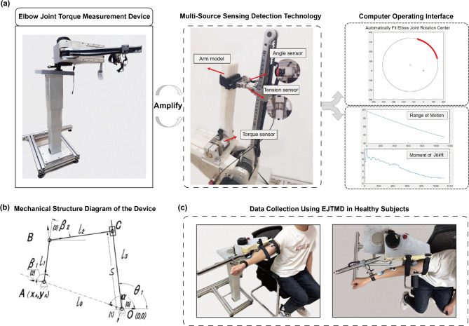
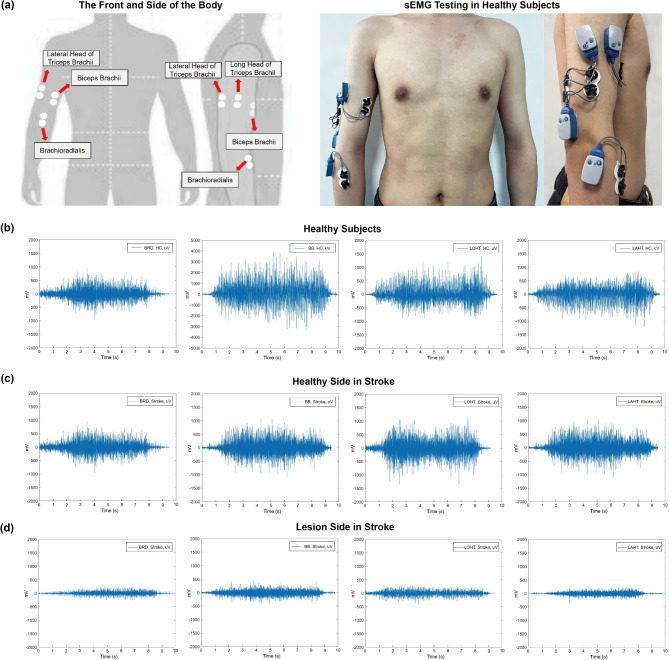
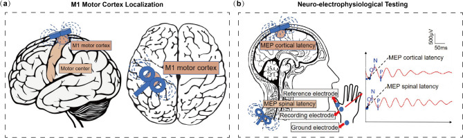
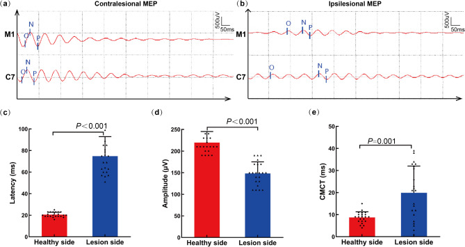
Current clinical practice lacks quantitative assessment methods for elbow joint movements. In response to existing research limitations, this study introduces the innovative elbow joint torque measurement device (EJTMD), which concurrently measures muscle strength and active range of motion (AROM) using a five-bar linkage system governed by a sliding mode control algorithm. Healthy subjects (n = 22) and stroke patients (n = 22) were recruited in a randomized trial. Each participant underwent two measurement. EJTMD or traditional tools like a protractor and a muscle strength tester. Participants were randomly allocated to EJTMD first or traditional tools first. The efficacy of EJTMD was assessed by comparing muscle strength and AROM with traditional tools. Integrated electromyography (iEMG) and root mean square (RMS) were used to assess the intensity of muscle activity during elbow movements. The peak torque (PT) and the ratio of peak torque to body weight (PT/BW) were examined to explore the differences in mechanical characteristics of bilateral elbow joints. Motor evoked potential (MEP) and central motor conduction time (CMCT) were employed to investigate the potential mechanisms underlying motor discrepancies post-stroke. EJTMD demonstrates superior muscle strength, AROM, iEMG, and RMS during elbow movements compared to traditional tools (P < 0.05). Repeated EJTMD measurement outcomes have a good correlation on the same day (r ≥ 0.999, P < 0.001). EJTMD exhibits significant differences in measurement outcomes among stroke patients before and after treatment (P < 0.05), compared to traditional tools. Stroke patients exhibit reduced PT and PT/BW on the lesion side across low, medium, and high-speed tests, with a more pronounced decline observed during low-speed testing (P < 0.001). Stroke patients show decreased iEMG and RMS on the affected side during elbow movements (P < 0.05), with prolonged MEP latency and CMCT (P < 0.001), and reduced MEP amplitude (P < 0.001). Based on the results, EJTMD demonstrates reliability and effectiveness in assessing elbow movements in both healthy subjects and stroke patients, showing sensitivity to minor joint changes. Stroke patients exhibit reduced flexor and extensor function on the lesion side, potentially resulting from impaired corticospinal tract conduction.
Keywords: Elbow joint; Healthy subjects; Motor evoked potential; Stroke; Surface electromyography.
© 2025. The Author(s).
Conflict of interest statement
Declarations. Competing interests: The authors declare no competing interests. Ethical approval: The research adhered to the reporting guidelines and regulations outlined in the Consolidated Standards of Reporting Trials (CONSORT) and the Declaration of Helsinki, approved by the Hospital Ethics Committee (application number: PJ2024-06-18), and was approved on 28/02/2023 by the Chinese Clinical Trial Registry (registration number: ChiCTR2300068710). All procedures were performed in accordance with the relevant guidelines and regulations.
Figures
References
-
- Zhang, W. et al. Examining the effectiveness of motor imagery combined with non-invasive brain stimulation for upper limb recovery in stroke patients: a systematic review and meta-analysis of randomized clinical trials. J. Neuroeng. Rehabil. 21, 209. 10.1186/s12984-024-01491-x (2024). - PMC - PubMed
-
- Hayward, K. S. et al. Timing and dose of upper limb motor intervention after stroke: a systematic review. Stroke52, 3706–3717. 10.1161/strokeaha.121.034348 (2021). - PubMed
-
- Gregory, W. J. & Saygin, D. Assessment of physical activity and muscle function in adult inflammatory myopathies. Curr. Rheumatol. Rep.24, 54–63. 10.1007/s11926-022-01059-5 (2022). - PubMed
-
- Juan, C., Xiang, C. & Minfen, S. A framework for daily activity monitoring and fall detection based on surface electromyography and accelerometer signals. IEEE J. Biomedical Health Inf.17, 38–45. 10.1109/titb.2012.2226905 (2013). - PubMed
Publication types
MeSH terms
Grants and funding
LinkOut - more resources
Full Text Sources
Medical
