. 2025 Apr 23;147(16):13699-13710.
doi: 10.1021/jacs.5c01337. Epub 2025 Apr 13.

Observation of the Assembly of the Nascent Mineral Core at the Nucleation Site of Human Mitochondrial Ferritin

Affiliations

Observation of the Assembly of the Nascent Mineral Core at the Nucleation Site of Human Mitochondrial Ferritin

Justin M Bradley et al. J Am Chem Soc. .

Abstract

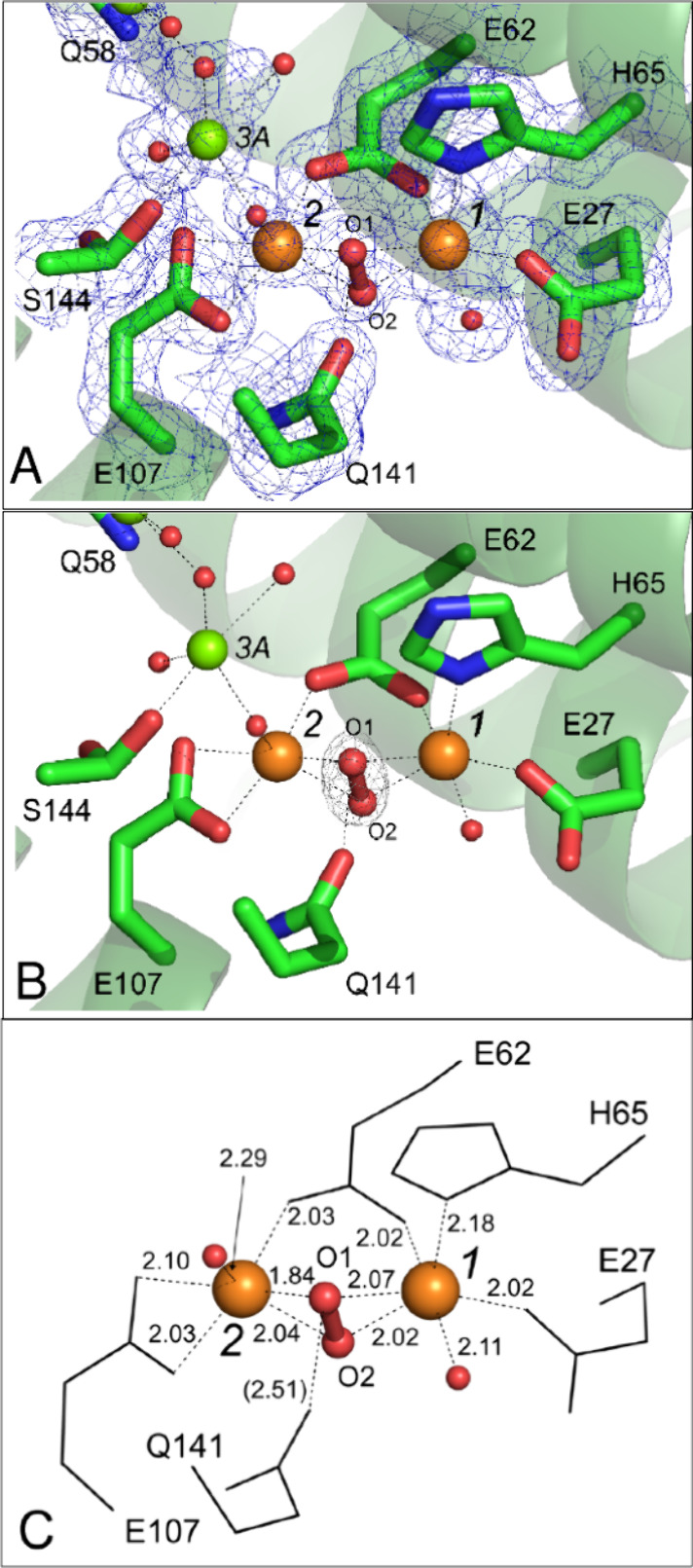
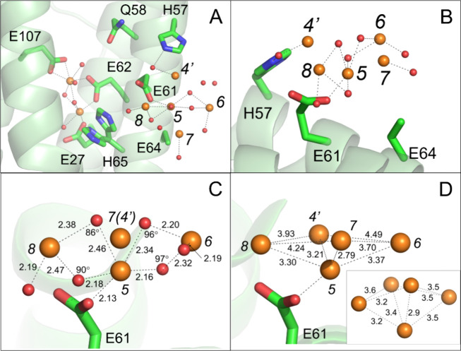
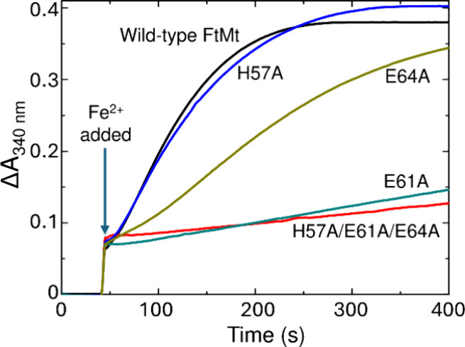
Ferritins play a crucial role in iron homeostasis and detoxification in organisms from all kingdoms of life. They are composed of 24 α-helical subunits arranged around an interior cavity where an iron-containing mineral core can be reversibly stored. Despite decades of study, leading to significant progress in defining the routes of Fe2+ uptake and the mechanism of its subsequent oxidation to Fe3+ at diiron catalytic sites termed ferroxidase centers, the process of core synthesis from the product of ferroxidase center activity remains poorly understood. In large part, this is due to the lack of high-resolution structural data on ferritin cores anchored to their nucleation sites on the inner surface of the protein. Mitochondrial ferritins are atypical of those found in higher eukaryotes in that they are homopolymers in which all subunits contain both a ferroxidase center and a presumed but undefined core nucleation site. Here, in conjunction with a novel method for producing iron-enriched ferritin crystals, we exploit these unusual features to structurally characterize both the nucleation site of mitochondrial ferritin and a pentanuclear, ferrihydrite-like iron-oxo cluster formed there. Kinetic data for wild-type and variant proteins confirmed the functional importance of this site, indicating a critical role for E61 in the transfer of Fe3+ from the ferroxidase center to the nascent mineral core.


Conflict of interest statement

The authors declare no competing financial interest.

Figures

Figure 1
Figure 1
Iron transport and mineralization in ferritins. (A) Fe2+ enters animal ferritin via the 3-fold channel, and is proposed to be shuttled to the FoC via transient binding at a series of sites (Fe3–Fe5) on the inner surface of the protein. (B) The Glu residues involved in Fe2+ coordination at these sites are structurally equivalent to Fe3+-coordinating residues in the ferric-oxo cluster anchored to the inner surface of HuLF. Images generated from PDBs 7o68 (FtMt) and 6tsf (HuLF). Irons and oxygens are shown as orange/brown and red spheres, respectively.
Figure 2
Figure 2
Exposure to O2 leads to formation of site of mineral core nucleation in iron-loaded FtMt. (A) Hydrated iron bound to site 4, a proposed transfer site involved in transport of Fe2+ substrate to the ferroxidase center in anaerobically harvested FtMt/Fe2+ cocrystals. The magenta mesh in this and panel (C) shows the anomalous difference Fourier map contoured at 6 σ. Metal binding sites are indicated by italicized black numbers. These include the locations of the proposed iron transfer sites 3A and 3B leading to the ferroxidase center occupied by magnesium ions in this structure. Iron positions are indicated by orange spheres, magnesium ions by green spheres, oxygen by red spheres. Metal coordination bonds are shown as black dashed lines. Individual residues coordinating the metal ions are shown in stick format and labeled. In this panel and panel (C) the minor conformer (28% occupancy) of His65 is not shown. (B) The equivalent structure derived from crystals anaerobically exposed to ferrous ammonium sulfate for 5 min (PDB entry 7O6A). (C) As (A) but for cocrystals exposed to aerobic well solution for 2 min prior to harvesting showing iron occupancies in sites 2 and 4 have decreased and peroxide is bound at the ferroxidase center. (D) The appearance of a large volume of anomalous difference electron density, modeled as an iron-oxo cluster in the vicinity of site 5 following exposure of cocrystals to aerobic well solution for 20 min prior to harvesting. Three iron sites (4’, 5 and 6) were found at peak heights at or above 4.0 σ in the anomalous difference electron density map (contoured at 4 σ in this panel). Site 4’ lies 1.8 Å from site 4 found in the structure of anaerobically harvested FtMt/Fe2+. Iron sites labeled 7 and 8 were located by MR-SAD and confirmed by ANODE. These are found at peak heights of 3.2 σ and 3.0 σ, respectively. Note that the side chain of residue E64 is presumed disordered as no significant electron density beyond atom Cγ was observed. In this panel a second conformer of His65 with significant occupancy (53%) is also shown.
Figure 3
Figure 3
Ferroxidase center in FtMt/Fe2+ cocrystals exposed to aerobic well solution for 2 min prior to harvest. (A) Blue mesh shows the Sigma-A weighted Fourier (2mFo-DFc) map contoured at 1.1 σ. Iron positions are indicated by orange spheres, oxygen by red spheres. Individual residues are shown in stick format and labeled. Metal-binding sites are indicated by italicized black numbers and metal coordination bonds are shown as black dashed lines. The oxygen atoms of the peroxide bridging the ferroxidase iron positions are labeled O1 and O2. (B) as (A) but gray mesh shows the peroxide omit map contoured at 6σ. (C) as (A) but showing detail of the geometry of the FoC. Individual residues are shown in wire format and labeled. The lengths of metal coordination bonds are shown in Ångstrom units. The length of the hydrogen bond from the side chain of Gln141 to peroxide atom O1 is indicated in brackets. Other geometry values are provided in Table 2.
Figure 4
Figure 4
An iron-oxo cluster on the inner surface of FtMt. (A, B) Approximately orthogonal views of an iron-oxo cluster in the vicinity of site 5 following exposure of cocrystals to aerobic well solution for 20 min prior to harvesting. Blue mesh shows the Sigma-A weighted Fourier (2mFo-DFc) map contoured at 1.3 σ within 4 Å of potential iron metal coordinating residues and the metal ions themselves. Iron positions are indicated by orange spheres, oxygen by red spheres. Individual residues are shown in stick format and labeled. Note that the side chain of residue E64 is presumed disordered as no significant electron density beyond atom Cγ was observed. Metal coordination bonds are shown as black dashed lines. (C), (D) As (A) but blue mesh shows the iron omit and oxygen omit maps, respectively, contoured at 3σ.
Figure 5
Figure 5
Iron-oxo cluster. (A) A view from inside the ferritin protein cage showing the ferroxidase center and iron-oxo cluster in the structure of FtMt observed following exposure of cocrystals to aerobic well solution for 20 min prior to harvesting. Iron positions are indicated by orange spheres, oxygen by red spheres. Individual residues coordinating the metal ions are shown in stick format and labeled. Metal binding sites are indicated by italicized black numbers and metal coordination bonds are shown as black dashed lines. Cartoon representation of FtMt polypeptide shown in green. (B) as (A) but showing an orthogonal close-up view of the iron-oxo cluster. (C) The geometry of the nascent mineral core. Interatomic distances where shown are in Ångstrom, angles in degrees. Note that in this view Fe4’ is occluded by Fe7. (D) Geometry of the cluster iron substructure. A putative platform for mineral core growth provided by iron sites 4’, 6, 7, and 8. Inset shows a comparable arrangement found within a proposed structure for ferrihydrite.
Figure 6
Figure 6
Iron mineralization by FtMt and inner surface variants. The increase in absorbance at 340 nm as a function of time following the aerobic addition of Fe2+ to a final concentration of 200 μm to 0.5 μm solutions of FtMt. The arrow indicates the point of Fe2+ addition.
Figure 7
Figure 7
Regeneration of rapid Fe2+ oxidation by FtMt. The increase in absorbance at 340 nm following the aerobic mixing of Fe2+ and FtMt either immediately following the oxidation of 200 equiv of Fe2+ (red) or after a further period of 3 (blue), 8 (yellow), 15 (green), 60 (cyan) or 1000 (orange) min. Data for (A) wild-type, (B) H57A/E61A/E64A, (C) H57A, (D) E61A or (E) E64A FtMt; fits to a biexponential decay are shown as black lines. Panel (F) shows the percentage of the rapid activity of the corresponding apo protein regenerated for time points up to 60 min for wild-type (black) or variant H57A/E61A/E64A (red), H57A (blue), E61A (dark cyan) or E64A (dark yellow) FtMt.
Figure 8
Figure 8
Coordinated side chain movements separate the Fe2+ uptake and Fe3+ release pathways in FtMt. Blue arrows indicate the route of Fe2+ entry into, and orange arrows the route of Fe3+ exit from, the FoC to the developing mineral core. Curved black arrows indicate the direction of side chain movement on Fe3+ release from the FoC. Movement of H65 triggers Fe3+ release from the FoC, with subsequent rearrangement of E61 to transport the product to site Fe5 on the inner surface for incorporation into the growing mineral core. Data following exposure of crystals to O2 for 20 min did not permit placement of the carboxylate group of E64 but the rotomer conformation is clearly different to that prior to cluster formation. We propose that this movement guides incoming Fe2+ away from the route of Fe3+ release and a breakdown in this separation is the origin of decreased mineralization activity in variant E64A.

Similar articles

References

    1. Bradley J. M.; Le Brun N. E.; Moore G. R. Ferritins: furnishing proteins with iron. J. Biol. Inorg. Chem. 2016, 21 (1), 13–28. 10.1007/s00775-016-1336-0. - DOI - PMC - PubMed
    1. Theil E. C. Ferritin protein nanocages use ion channels, catalytic sites, and nucleation channels to manage iron/oxygen chemistry. Curr. Opin Chem. Biol. 2011, 15 (2), 304–311. 10.1016/j.cbpa.2011.01.004. - DOI - PMC - PubMed
    1. Theil E. C.; Behera R. K.; Tosha T. Ferritins for chemistry and for life. Coord Chem. Revs 2013, 257 (2), 579–586. 10.1016/j.ccr.2012.05.013. - DOI - PMC - PubMed
    1. Crichton R. R.; Declercq J. P. X-ray structures of ferritins and related proteins. Biochim. Biophys. Acta 2010, 1800 (8), 706–718. 10.1016/j.bbagen.2010.03.019. - DOI - PubMed
    1. Briat J. F.; Duc C.; Ravet K.; Gaymard F. Ferritins and iron storage in plants. Biochim. Biophys. Acta 2010, 1800 (8), 806–814. 10.1016/j.bbagen.2009.12.003. - DOI - PubMed