

Roots Form and Canals Morphology of Maxillary Second Premolar in a Sample of Yemeni Population
- PMID: 40223865
- PMCID: PMC11986953
- DOI: 10.1155/ijod/3380604
Roots Form and Canals Morphology of Maxillary Second Premolar in a Sample of Yemeni Population
Abstract
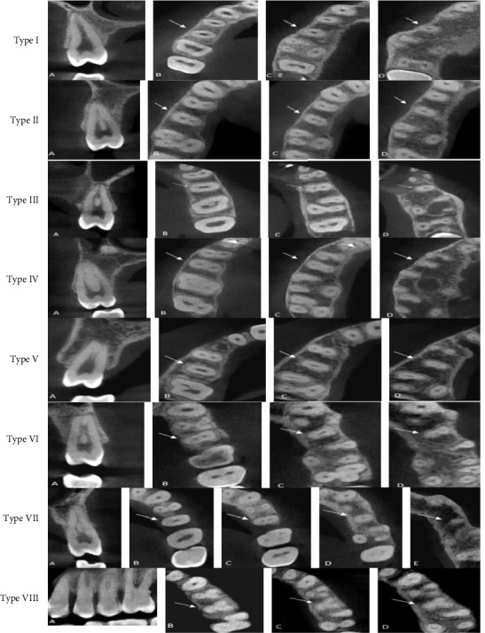
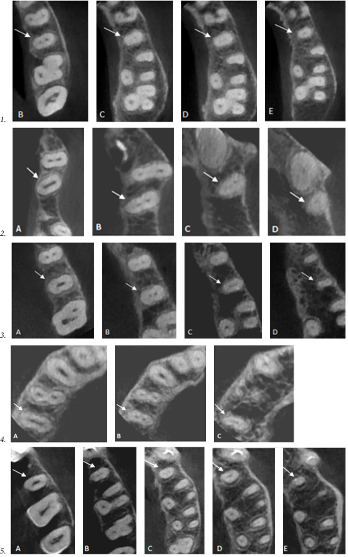
Background: The present study aimed to investigate the root form and canal anatomy of the permanent maxillary second premolar in a sample of Yemeni population using cone beam computed tomography (CBCT). Methods: A total of 362 CBCT scans of maxillary second premolars of Yemeni patients aged between 15 and 60 years were analyzed to determine the anatomy of this tooth including number and form of roots, number of canals, number of orifices, and root canal configurations. Chi-square test was used to analyze the association between different variables. Results: Of the 362 examined maxillary second premolars, 87.6% had one root, 12.1% had two roots, and 0.3% had three fused roots. Regarding the canal number, one canal was found in 181 teeth (50%), while two canals were found in 180 teeth (49.7%), three canals were found in one tooth (0.3%). One orifice was observed in 263 teeth (72.7%), two orifices in 98 teeth (27.1%), and three orifices were reported in one tooth (0.3%). Regarding root canal configuration, 98.6% of the sample were within the eight types of Vertucci classification. The majority of single-rooted second premolars (46.1%) exhibited type I canal configuration, type III found in 14.1%, and type II found in 9.9%. However, type IV found in 13.2% (48 teeth) of the sample, out of these, 88.6% (39 teeth) had two roots. Type VIII was observed in the three-rooted tooth (0.3%). Supplemental and additional canal types were found in 1.1% of the sample. Moreover, a new canal type was observed in 0.3% of the sample. According to Ahmed's coding system the most prevalent type was 1MSP 1-1 (46.1%) followed by 1MSP1-2-1 (4.1%), then type 2MSP B1 P1 (10.8%). Chi-square tests showed that the difference in root canal configurations among male and female was statistically significant. Conclusions: Root and canal morphology of maxillary second premolars among the evaluated Yemeni population is highly variable and requires cautious evaluation prior to endodontic treatment. Majority of the sample were single-rooted teeth, most of them had a complicated and variable canal configuration. Moreover, significant gender disparities in internal and external morphology were observed.
Keywords: Yemeni population; configuration; endodontics; maxillary second premolar; root canal morphology.
Copyright © 2025 AmatAlkhaliq M. Al-Sayaghi et al. International Journal of Dentistry published by John Wiley & Sons Ltd.
Conflict of interest statement
The authors declare no conflicts of interest.
Figures


Similar articles
-
The morphology of permanent maxillary first molars evaluated by cone-beam computed tomography among a Yemeni population.BMC Oral Health. 2023 Jan 27;23(1):46. doi: 10.1186/s12903-023-02752-2. BMC Oral Health. 2023. PMID: 36703140 Free PMC article.
-
A cone-beam computed tomography study of root canal morphology of maxillary and mandibular premolars in a Turkish population.Acta Odontol Scand. 2014 Nov;72(8):701-6. doi: 10.3109/00016357.2014.898091. Epub 2014 May 15. Acta Odontol Scand. 2014. PMID: 24832561
-
Morphological Evaluation of Maxillary Premolar Canals in Iranian Population: A Cone-Beam Computed Tomography Study.J Dent (Shiraz). 2020 Sep;21(3):215-224. doi: 10.30476/DENTJODS.2020.82299.1011. J Dent (Shiraz). 2020. PMID: 33062816 Free PMC article.
-
Anatomical Evaluation of Maxillary Premolars in a Saudi Population: An In Vivo Cone-beam Computed Tomography Study.J Contemp Dent Pract. 2021 Mar 1;22(3):284-289. J Contemp Dent Pract. 2021. PMID: 34210930
-
Cone-beam Computed Tomography Analysis of the Root Canal Morphology of Maxillary First and Second Premolars in a Spanish Population.J Endod. 2015 Aug;41(8):1241-7. doi: 10.1016/j.joen.2015.03.026. Epub 2015 May 5. J Endod. 2015. PMID: 25956606
References
-
- Vertucci F. J. Root Canal Morphology and Its Relationship to Endodontic Procedure. Endodontic Topics . 2005;10(1):3–29.
-
- Pécora J. D., Sousa Neto M. D., Saquy P. C., Woelfel J. B. In Vitro Study of Root Canal Anatomy of Maxillary Second Premolars. Brazilian Dental journal . 1993;3(2):81–85. - PubMed
LinkOut - more resources
Full Text Sources
Research Materials
