. 2025 Mar 15:7:100157.
doi: 10.1016/j.bbadva.2025.100157. eCollection 2025.

Evolution and function of galectins in Xenopus laevis: Comparison with mammals and new perspectives

Affiliations

Evolution and function of galectins in Xenopus laevis: Comparison with mammals and new perspectives

Takashi Ogawa et al. BBA Adv. .

Abstract

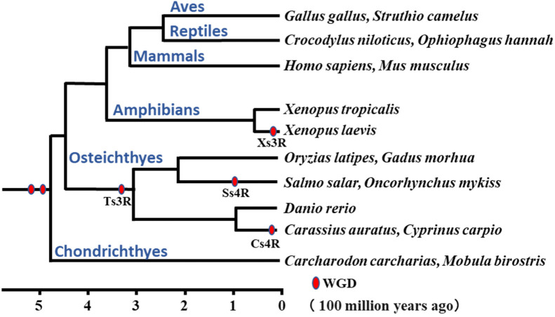
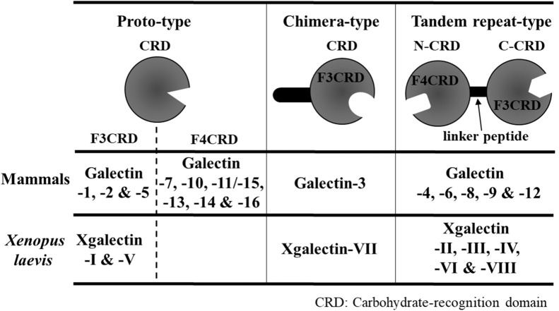
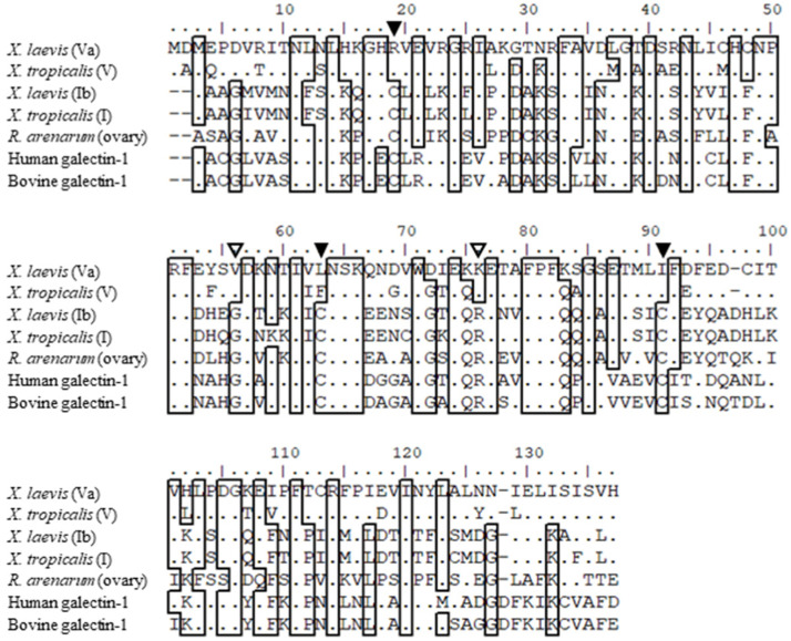
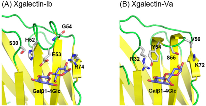
Galectins are metal-independent sugar-binding proteins that recognize galactose (the β-galactoside structure) and regulate the cross-linking of sugar chains between cells and the extracellular matrix. Their specificity for galactose is attributed to their highly conserved carbohydrate recognition domain. Galectins participate in biological processes across species, including development, differentiation, morphogenesis, tumor progression, metastasis, immunity, and apoptosis. However, the relationship between the binding of galectin to sugar chains (glycans) and their biological functions remains unclear. Thus, a comprehensive functional analysis of galectins is required to better characterize their evolutionarily conserved and unique functions. We have previously identified and characterized 12 Xenopus laevis galectins (xgalectins), the only non-mammalian vertebrate species in which galectins have been comprehensively characterized to date. In this review, we present the latest findings on the types and functions of xgalectins and discuss prospects for elucidating their diverse functions from an evolutionary perspective through comparisons with mammalian galectins.

Keywords: Carbohydrate recognition; Evolution; Galectins; Sequence conservation; Whole-genome duplication; Xenopus laevis.


Conflict of interest statement

The authors declare that they have no known competing financial interests or personal relationships that could have appeared to influence the work reported in this paper.

Figures

Fig 1
Fig. 1
Phylogenetic tree of vertebrates with whole genome duplication events. Estimated timing of whole genome duplication (WGD) events in vertebrate phylogeny and evolution. The phylogenetic tree is shown with divergent ages of taxa and representative animals listed on the right. WGD events (indicated by asterisks) occurred twice in the common ancestor of vertebrates approximately 500 million years ago. In teleost fish, the third WGD (Ts3R) occurred approximately 320 million years ago, followed by a forth WGD about 100 and 15 million years ago in the salmonidae lineage (Ss4R) and cyprinidae lineage (Cs4R). In amphibians, the third WGD occurred 18 million years ago in the lineage of Xenopus laevis (Xs3R).
Fig 2
Fig. 2
Classification of mammalian and Xenopus laevis galectins based on structure. Proto- and chimera-type galectins contain one carbohydrate recognition domain (CRD), whereas tandem-repeat galectins possess two CRDs. Each CRD comprises approximately 140 amino acids and is classified as either F3CRD or F4CRD. The 7 amino acid residues important for sugar recognition are conserved in most galectins across species. Proto- and chimera-type galectins have only one CRD but can bind to multiple carbohydrate chains by dimer or polymer formation, depending on the molecular species of galectin. N—CRD, N-terminal CRD; C—CRD, C-terminal CRD.
Fig 3
Fig. 3
Amino acid sequence alignment of proto-type galectins. Sequences were aligned using ClustalW (gap opening penalty was 10, gap extension penalty was 0.05, and Gonet protein weight matrix was used). Amino acid residues conserved in five or more sequences are highlighted with frames. Amino acids conserved in xgalectin-Va are shown as dots. Cysteine residues conserved in galectin-1 homologs are marked by closed triangles. Amino acids mentioned in the manuscript as being involved in ligand selectivity are marked by open triangles.
Fig 4
Fig. 4
Amino-acid residues involved in carbohydrate binding selectivity. Residues involved in ligand selectivity and ligand molecules are represented as stick models. Although Gly54 (Val56) is distant from the ligand, it affects the conformation of the loop region. (A) Structure of xgalectin-Ib/lactose complex. (B) Structure of galectin-Va/lactose complex.
Fig 5
Fig. 5
Crystal structures of Rhinella arenarum and Xenopus laevis proto-type galectins (A) Structure of R. arenarum ovary galectin dimer. (B) Structure of xgalectin-Ib dimer, which resembles the dimer structures of toad and mammalian galectin-1s. (C) Structure of xgalectin-Va tetramer. Two xgalectin-1-like dimers (green/cyan and magenta/yellow) assemble into a tetramer.
Fig 6
Fig. 6
Amino acid sequence alignment of xgalectin-VIIa, -VIIb, and human galectin-3. Sequences were aligned and the repeat units (I-V) are underlined according to cooper et al., [93]. The black line indicates the human repeating unit, and the red line indicates the repeating unit characteristic of xgalectin-VIIb. Gaps are represented by hyphens. Two amino acid substitutions are indicated with arrow heads (K176 M and N180 K).

Similar articles

References

    1. Ohno S., Wolf U., Atkin N.B. Evolution from fish to mammals by gene duplication. Hereditas. 1968;59:169–187. doi: 10.1111/j.1601-5223.1968.tb02169.x. - DOI - PubMed
    1. Van de Peer Y., Maere S., Meyer A. The evolutionary significance of ancient genome duplications. Nat. Rev. Genet. 2009;10:725–732. doi: 10.1038/nrg2600. - DOI - PubMed
    1. S. Ohno, Evolution by gene duplication (1970).
    1. Holland P.W., Garcia-Fernandez J., Williams N.A., Sidow A. Gene duplications and the origins of vertebrate development. Dev. Suppl. 1994:125–133. - PubMed
    1. Houzelstein D., Goncalves I.R., Fadden A.J., Sidhu S.S., Cooper D.N., Drickamer K., Leffler H., Poirier F. Phylogenetic analysis of the vertebrate galectin family. Mol. Biol. Evol. 2004;21:1177–1187. doi: 10.1093/molbev/msh082. - DOI - PubMed

LinkOut - more resources