

Optimization and Validation of the Cationization Method for the Fab' Fragment of Antibody Rituximab
- PMID: 40224423
- PMCID: PMC11983203
- DOI: 10.1021/acsomega.4c07727
Optimization and Validation of the Cationization Method for the Fab' Fragment of Antibody Rituximab
Abstract
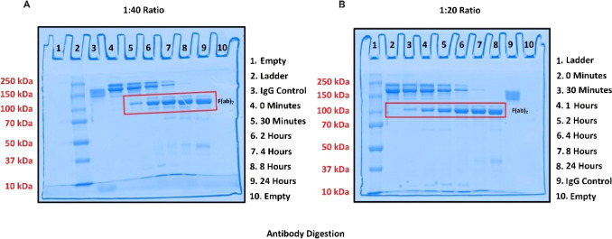
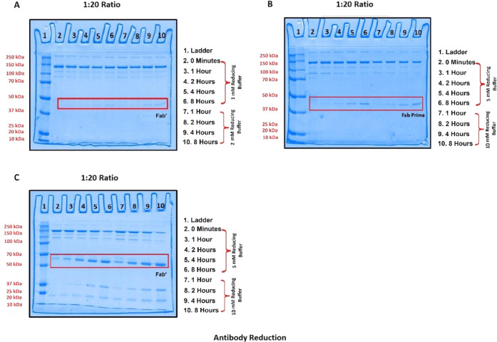
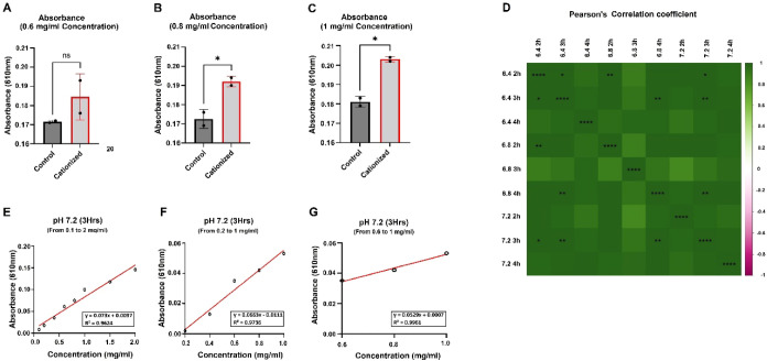
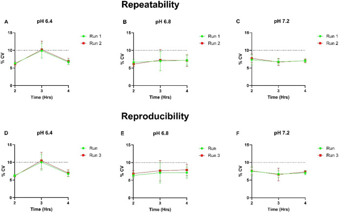
This study aimed to optimize and validate a cationization method for the Fab' fragment of antibody rituximab, following the guidelines set by the International Council for Harmonization of Technical Requirements for Pharmaceuticals for Human Use (ICH). The optimization process involved fragmentation of antibody rituximab into an F(ab)2 fragment using pepsin, followed by reduction into the Fab' fragment, and subsequent cationization. Various parameters, such as time intervals, enzyme ratios, reducing agent concentrations, pH levels, and reaction durations, were systematically investigated to achieve optimal cationization efficiency. The developed method was validated through spectrophotometry using the bromophenol blue (BPB) dye assay method. The validation process included assessment of linearity, robustness, and sensitivity. Results demonstrated the efficacy of the optimized cationization method, providing a reliable approach for analyzing rituximab antibody cationization of Fab'.
© 2025 The Authors. Published by American Chemical Society.
Conflict of interest statement
The authors declare no competing financial interest.
Figures
Similar articles
-
Pretargeting for cancer radioimmunotherapy with bispecific antibodies: role of the bispecific antibody's valency for the tumor target antigen.Bioconjug Chem. 2002 Sep-Oct;13(5):1054-70. doi: 10.1021/bc0200172. Bioconjug Chem. 2002. PMID: 12236788
-
Monoclonal antibody Fab' fragment cross-linking using equilibrium transfer alkylation reagents. A strategy for site-specific conjugation of diagnostic and therapeutic agents with F(ab')2 fragments.Bioconjug Chem. 1994 May-Jun;5(3):220-35. doi: 10.1021/bc00027a007. Bioconjug Chem. 1994. PMID: 7918742
-
[High level expression of chimeric antibody fragment F(ab')2 directed against CD20 in Escherichia coli].Sheng Wu Gong Cheng Xue Bao. 2004 Sep;20(5):673-8. Sheng Wu Gong Cheng Xue Bao. 2004. PMID: 15973988 Chinese.
-
Site-specific conjugation of bifunctional chelator BAT to mouse IgG1 Fab' fragment.Acta Pharmacol Sin. 2006 Feb;27(2):237-41. doi: 10.1111/j.1745-7254.2006.00242.x. Acta Pharmacol Sin. 2006. PMID: 16412275
-
Improvement of therapeutic effect by using Fab' fragment in the treatment of carcinoembryonic antigen-positive human solid tumors with adriamycin-entrapped immunoliposomes.Jpn J Cancer Res. 1994 Apr;85(4):434-40. doi: 10.1111/j.1349-7006.1994.tb02377.x. Jpn J Cancer Res. 1994. PMID: 8200855 Free PMC article.
References
-
- Raso V. Antibodies in diagnosis and therapy. The magic bullet--nearing the century mark. Semin. Cancer Biol 1990, 1 (3), 227–242. - PubMed
LinkOut - more resources
Full Text Sources