. 2025 Mar 27;10(13):13570-13582.
doi: 10.1021/acsomega.5c00476. eCollection 2025 Apr 8.

Enhanced Ear Cartilage Regeneration with Dual-Network LT-GelMA/F127DA Hydrogel Featuring Nanomicelle Integration

Affiliations

Enhanced Ear Cartilage Regeneration with Dual-Network LT-GelMA/F127DA Hydrogel Featuring Nanomicelle Integration

Bingzhang Liu et al. ACS Omega. .

Abstract

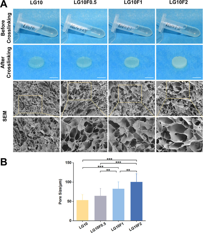
Tissue-engineered cartilage, supported by advancements in photo-cross-linkable hydrogels, offers a promising solution for the repair and regeneration of damaged cartilage in anatomically complex and mechanically demanding sites. Low-temperature soluble GelMA (LT-GelMA) remains in a liquid state at room temperature, allowing for easier handling; however, it has limitations in mechanical strength and structural stability. To address these limitations, we developed a novel dual-network hydrogel combining LT-GelMA with Pluronic F127-diacrylate (F127DA). The resulting hydrogel uniquely integrates the low-temperature solubility of LT-GelMA with the enhanced mechanical strength provided by photo-cross-linkable F127DA nanomicelles. Additionally, the hydrogel exhibits controlled swelling and biodegradation rates. In vitro studies revealed a significant increase in chondrocyte viability by day 7 in formulations with higher F127DA concentrations. In vivo, the hydrogel demonstrated superior neo-cartilage formation in a subcutaneous nude mouse model, as indicated by increased deposition of cartilage-specific extracellular matrix components at 4 and 8 weeks. In summary, we developed a hydrogel with fluidity at room temperature and enhanced mechanical performance. These results indicate that the LT-GelMA/F127DA hydrogel effectively addresses the current gaps in cartilage tissue engineering. The hydrogel's superior performance, especially in promoting cartilage regeneration, positions it as a promising alternative for reconstructive surgery, representing a significant improvement over existing cartilage repair strategies.


Conflict of interest statement

The authors declare no competing financial interest.

Figures

Figure 1
Figure 1
Schematic of LT-GelMA/F127DA bioink preparation and its application in cartilage regeneration. F127DA was incorporated into LT-GelMA to create a premixed solution. Chondrocytes were isolated and expanded from rabbit ears, then mixed with the LT-GelMA/F127DA premixture to prepare the bioink. The bioink was photo-cross-linked using a 405 nm curing light to form a dual-network structure in the shape of disk-like scaffolds. These scaffolds were subsequently cultured in vitro for cell viability assays and implanted subcutaneously into nude mice for in vivo experiments.
Figure 2
Figure 2
Schematic illustration of the preparation and cross-linking process of LT-GelMA/F127DA hydrogels. F127DA (represented by a two-color wavy line) self-assembles into nanomicelles (depicted as two-color sphere with red hydrophobic core and blue hydrophilic shell) in the hydrogel solution. Upon the addition of the photoinitiator LAP (shown as a purple ball) and exposure to 405 nm light, these micelles integrate with LT-GelMA (illustrated by a blue spiral) to form bonds (represented by purple dashed lines). The cross-linking process involves three critical interactions: micellar cross-linkings within F127DA nanomicelles, bridge linkages between F127DA nanomicelles and LT-GelMA, and direct covalent cross-linkings within LT-GelMA molecules. These interactions establish a robust dual-network structure.
Figure 3
Figure 3
Morphology of the bioinks with different concentrations of F127DA. (A) The optical images of the bioinks before cross-linking show no significant differences in color or transparency. The gross appearance after cross-linking showed that the transmittance decreased with higher concentration of F127DA (scale bar: 5 mm). The SEM images provide a detailed view of the cross-linked LT-GelMA/F127DA hydrogels. At low magnification (scale bar: 100 μm), the overall porous structure is evident, with increased pore uniformity and size at higher F127DA concentrations. At high magnification (scale bar: 50 μm), the images reveal the enlargement of the pore size as F127DA concentration increases. (B) Quantitative analysis using ImageJ demonstrates a statistically significant increase in average pore size with rising F127DA concentrations (*p < 0.05, **p < 0.01, ***p < 0.001).
Figure 4
Figure 4
Characterization of LT-GelMA/F127DA bioinks with varying F127DA concentrations. Different colors or lines correspond to bioinks with varying F127A concentrations. (A) Stress–strain curves: This panel illustrates the stress–strain behavior of the bioinks under compressive loading, revealing that the compressive modulus increases with higher F127DA concentrations. The curve’s shape provides insight into the material’s mechanical strength and elasticity, where steeper slopes indicate higher stiffness. (B) Compressive modulus: The compressive modulus, calculated from the linear region of the stress–strain curves, is plotted here as a bar graph. The compressive modulus increases significantly as the concentration of F127DA increases, demonstrating the enhancement of mechanical stiffness due to F127DA incorporation. (C) Viscosities of precuring bioinks: This subplot presents the viscosity of the bioinks before curing, measured as a function of shear rate. The curves show that bioinks with higher F127DA concentrations exhibit increased viscosity, maintaining stability over time. A lower viscosity indicates a thinner, less resistant bioink to flow. (D) Storage (G′) and loss (G″) modulus: the storage modulus (G′) and loss modulus (G″) of the bioinks are plotted as a function of angular frequency. G′ represents the elastic behavior and G″ indicates the viscous behavior. Both moduli increase as the F127DA concentration rises. A higher G′ compared to G″ suggests a more solid-like material, which is beneficial for structural stability postcuring. (E) Swelling ratio: This bar graph illustrates the swelling ratio of the hydrogels after immersion in PBS for a specified time. The swelling ratios of the cross-linked bioinks decrease inversely with increasing F127DA concentrations, highlighting improved structural integrity. (F) In vitro degradation: The remaining mass of the hydrogels is depicted as a function of time. The biodegradation profile in PBS solution reveals a slower degradation rate with higher F127DA concentrations, demonstrating enhanced longevity and stability (*p < 0.05, **p < 0.01, ***p < 0.001).
Figure 5
Figure 5
Results of in vitro culture of chondrocytes within LT-GelMA/F127DA bioink-based photo-cross-linked constructs. (A) 3D reconstructed confocal fluorescence images show the constructs at days 1 and 7. Live/Dead staining was performed to assess cell viability (scale bar: 500 μm). Green fluorescence indicates live cells, while red fluorescence marks dead cells. The images reveal high cell viability at day 1, with minimal red staining, and a significant increase in viability with increasing concentrations of F127DA at day 7. (B) Quantitative analysis of chondrocyte viability confirms the observations, showing high rates (>95%) at day 1 for all samples and a statistically significant increase at day 7, with constructs containing F127DA outperforming those with only LT-GelMA (*p < 0.05, **p < 0.01, ***p < 0.001).
Figure 6
Figure 6
In vivo neocartilage formation. (A) Top: picture taken right after the subcutaneous implantation of auricular. Bottom: picture taken during explantation of constructs after 8 weeks in vivo. (B) Images of in vivo constructs at weeks 4 and 8 (scale bar: 2 mm). (C) In vivo histological analysis for the development of cartilage tissue formation at 4 and 8 weeks after transplantation. Histological and immunohistological analyses for H&E, Safranin O, Toluidine Blue and Collagen II at low magnification (scale bar: 1 mm) and at high magnification (scale bar: 100 μm). (D) Collagen II positive staining area analysis of chondrocytes in constructs at weeks 4 and 8 (*p < 0.05, **p < 0.01, ***p < 0.001).

Similar articles

References

    1. Visscher D. O.; Lee H.; Van Zuijlen P. P. M.; Helder M. N.; Atala A.; Yoo J. J.; Lee S. J. A Photo-Crosslinkable Cartilage-Derived Extracellular Matrix Bioink for Auricular Cartilage Tissue Engineering. Acta Biomater. 2021, 121, 193–203. 10.1016/j.actbio.2020.11.029. - DOI - PMC - PubMed
    1. Song X.; Zhang P.; Luo B.; Li K.; Liu Y.; Wang S.; Wang Q.; Huang J.; Qin X.; Zhang Y.; Zhou G.; Lei D. Multi-Tissue Integrated Tissue-Engineered Trachea Regeneration Based on 3D Printed Bioelastomer Scaffolds. Advanced Science 2024, 11 (39), 2405420.10.1002/advs.202405420. - DOI - PMC - PubMed
    1. Pang L.; Jin H.; Lu Z.; Xie F.; Shen H.; Li X.; Zhang X.; Jiang X.; Wu L.; Zhang M.; Zhang T.; Zhai Y.; Zhang Y.; Guan H.; Su J.; Li M.; Gao J. Treatment with Mesenchymal Stem Cell-Derived Nanovesicle-Containing Gelatin Methacryloyl Hydrogels Alleviates Osteoarthritis by Modulating Chondrogenesis and Macrophage Polarization. Adv. Healthcare Mater. 2023, 12 (17), 2300315.10.1002/adhm.202300315. - DOI - PubMed
    1. Browe D. C.; Mahon O. R.; Díaz-Payno P. J.; Cassidy N.; Dudurych I.; Dunne A.; Buckley C. T.; Kelly D. J. Glyoxal Cross-linking of Solubilized Extracellular Matrix to Produce Highly Porous, Elastic, and Chondro-permissive Scaffolds for Orthopedic Tissue Engineering. J. Biomed. Mater. Res 2019, 107 (10), 2222–2234. 10.1002/jbm.a.36731. - DOI - PubMed
    1. Feng B.; Ji T.; Wang X.; Fu W.; Ye L.; Zhang H.; Li F. Engineering Cartilage Tissue Based on Cartilage-Derived Extracellular Matrix cECM/PCL Hybrid Nanofibrous Scaffold. Mater. Des. 2020, 193, 108773.10.1016/j.matdes.2020.108773. - DOI

LinkOut - more resources