. 2025 Mar 26;10(13):13105-13121.
doi: 10.1021/acsomega.4c10575. eCollection 2025 Apr 8.

Xuanfei Baidu Decoction Alleviated Sepsis-Induced ALI by Modulating Gut Microbial Homeostasis and Promoting Inflammation Resolution: Bioinformatics and Experimental Study

Affiliations

Xuanfei Baidu Decoction Alleviated Sepsis-Induced ALI by Modulating Gut Microbial Homeostasis and Promoting Inflammation Resolution: Bioinformatics and Experimental Study

Lei Yang et al. ACS Omega. .

Abstract

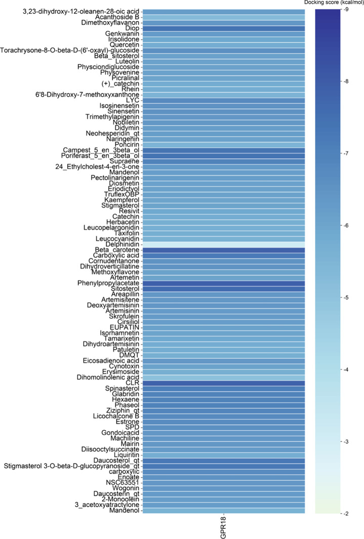
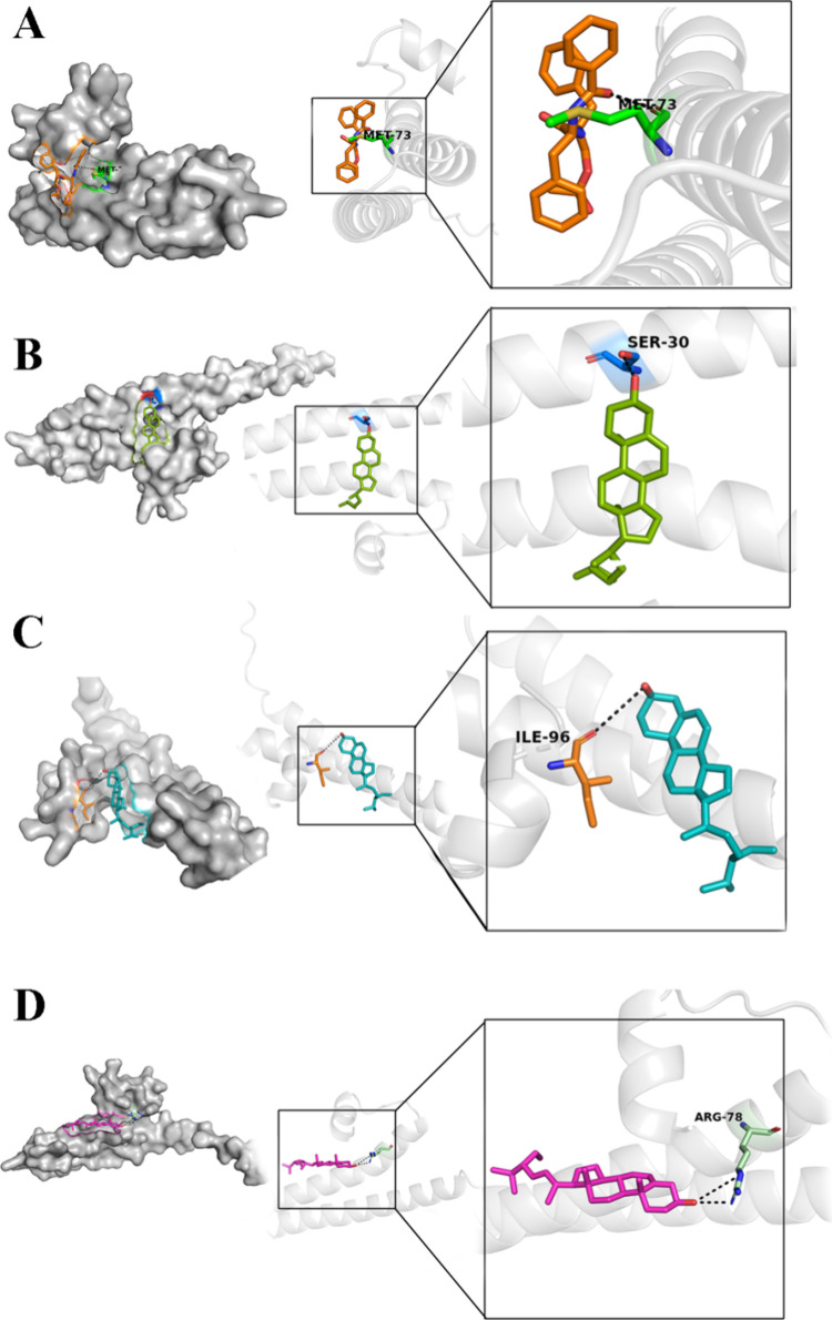
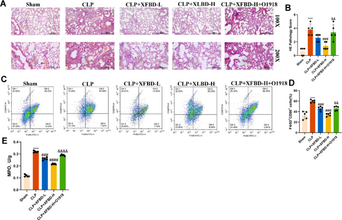
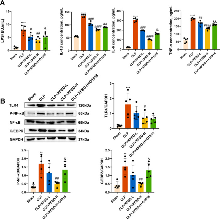
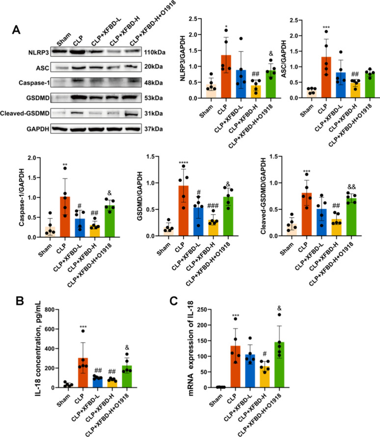
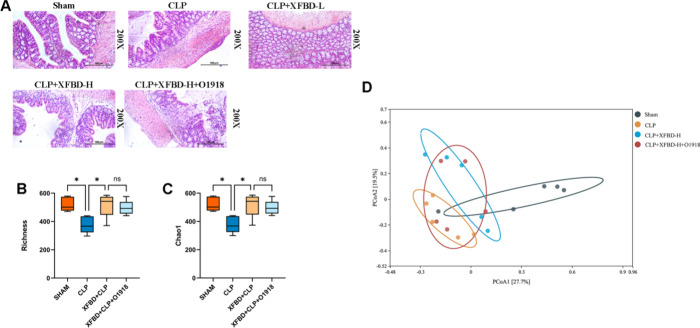
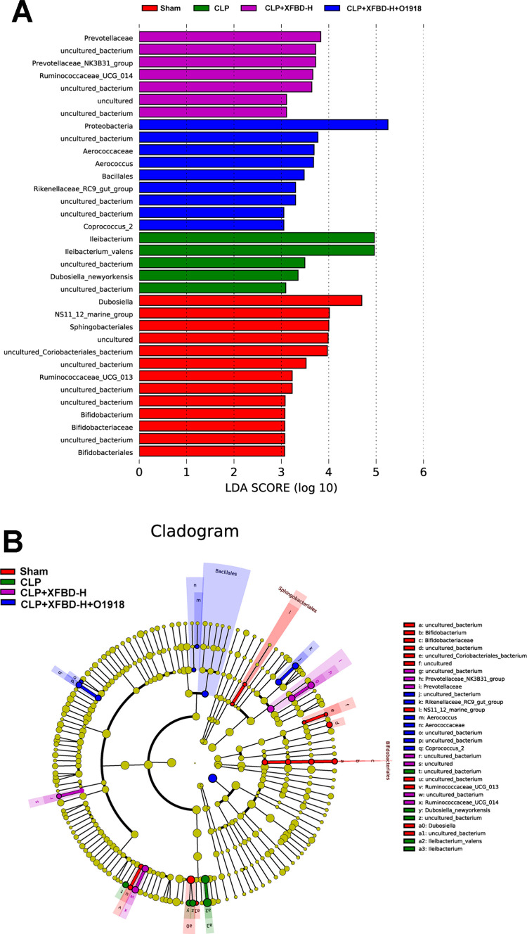
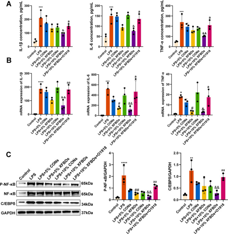
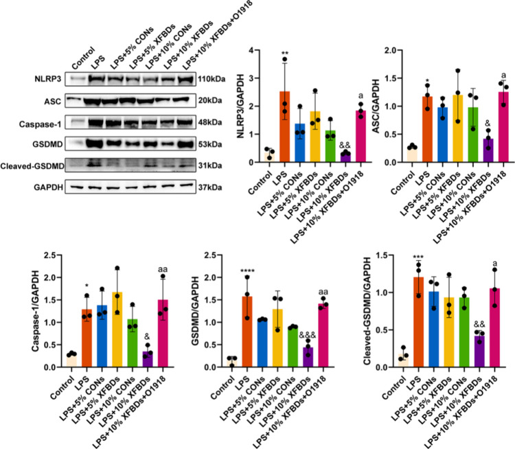
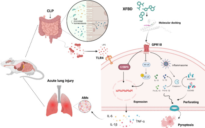
The Xuanfei Baidu Decoction (XFBD) has shown effective therapeutic potential for acute lung injury (ALI) induced by lipopolysaccharide and immunoglobin G immune complexes. Herein, the protective effects and mechanisms of XFBD were investigated in a sepsis-induced ALI mouse model along with its effects on gut microbiota. Notably, bioinformatics and molecular docking analyses revealed that XFBD components exhibited a strong binding affinity to G-protein-coupled receptor 18 (GPR18). In the murine ALI model-induced by cecal ligation and puncture (CLP)-XFBD markedly improved lung histopathology, reduced M1 macrophage polarization, and decreased pro-inflammatory cytokine levels in both lung tissues and MH-S macrophages. Furthermore, XFBD downregulated key inflammatory pathways, including nuclear factor (NF)-κB, phosphorylated-NF-κB, CCAAT/enhancer binding protein-δ, and the nucleotide-binding oligomerization domain-like receptor pyrin domain-containing 3/Caspase-1/gasdermin D axis. Additionally, XFBD restored the CLP-induced disruption in gut microbiota balance, increasing the abundance of Prevotellaceae and Ruminococcaceae_UCG_014. Altogether, the findings of this study suggest that XFBD alleviates CLP-induced ALI by modulating gut microbial homeostasis and inhibiting associated inflammatory pathways, particularly via GPR18 activation, presenting the promising therapeutic potential of XFBD for treating sepsis-induced ALI.


Conflict of interest statement

The authors declare no competing financial interest.

Figures

Figure 1
Figure 1
Top 50 degree-ranked targets and molecular docking results with GPR18.
Figure 2
Figure 2
Molecular docking results of the top four ranked docking scores: (A) 3-phenylpropyl acetate docking with GPR18; (B) beta-carotene docking with GPR18; (C) cholesterol docking with GPR18; (D) sitosterol docking with GPR18.
Figure 3
Figure 3
Results of XFBD component analysis by HPLC. (A) Chromatogram of a standard mixture XFBD, peak 1: amygdalin, peak2: verbenalin, peak 3: naringin, peak 4: glycyrrhetinic acid. (B) XFBD components, peak 1: amygdalin, peak2: verbenalin, peak 3: naringin, peak 4: glycyrrhetinic acid.
Figure 4
Figure 4
XFBD alleviated pathological damage in a mouse model of ALI induced by CLP, and the GPR18 inhibitor eliminated the anti-inflammatory response. (A) Lung tissue histopathological damage 24 h after CLP modeling evaluated by H&E staining, with images captured at 200× magnification. (B) Lung histopathology scores. (C) Proportion of F4/80+ CD86+ cells detected by flow cytometry in the spleens of CLP mouse models treated with PBS (control) or XFBD decoction. (D) The effect of XFBD on the MPO expression in the mouse model induced by CLP. All image data shown are representative of the experimental results. ****P < 0.0001 compared with the Sham group, ###P < 0.001 compared with the CLP group, &&P < 0.01 compared with the CLP + XFBD-H group.
Figure 5
Figure 5
Alleviating effect of the selective GPR18 agonist XFBD on CLP-induced ALI mice. (A) Levels of LPS, TNF-α, IL-1β, and IL-6 in peripheral blood were detected by ELISA. (B) Western blotting exhibits the expression of TLR4, P-NF-κB, NF-κB, and C/EBPδ proteins in the lung tissues. The data presented in Figure 9A,B represent the mean ± SEM of five independent experiments. **P < 0.01 when compared with the Sham group, ****P < 0.0001 when compared with the Sham group, ##P < 0.01 when compared with the CLP group, ###P < 0.001 when compared with the CLP group, ####P < 0.001 when compared with the CLP group, &P < 0.05 when compared with the CLP + XFBD-H group, &&P < 0.01 when compared with the CLP + XFBD-H group.
Figure 6
Figure 6
XFBD demonstrates the inhibition of GSDMD-mediated pyroptosis in ALI mice induced by IgG-IC, and the GPR18 inhibitor O1918 counteracted the antipyroptosis effect. (A) NLRP3, ASC, GSDMD, Cleaved-GSDMD, and Caspase-1 were detected by Western blotting. (B) ELISA detection of IL-18 concentrations in the serum. (C) qPCR assay detection of the IL-18 mRNA expressions in the lung tissues. Data are presented as the mean ± SEM for five independent experiments. *P < 0.05 when compared with the Sham group, **P < 0.01 when compared with the Sham group, ***P < 0.001 when compared with the Sham group, ****P < 0.0001 when compared with the Sham group, #P < 0.05 when compared with the CLP group, ##P < 0.01 when compared with the CLP group, ###P < 0.001 when compared with the CLP group, &P < 0.05 when compared with the CLP + XFBD-H group, &&P < 0.01 when compared with the CLP + XFBD-H group.
Figure 7
Figure 7
Effect of XFBD on the intestinal barrier and gut microbiota in mice with CLP-induced ALI. (A) Histopathological images of the colon tissues in each group. (B, C) Alpha diversity is illustrated using chao1 and Richness. (D) PCoA was used to analyze the bacterial communities of each group of β-diversity assessment. The significance of differences between the groups was evaluated using nonparametric factors Kruskal–Wallis and rank tests. *P < 0.05 is considered to indicate significant differences between the groups.
Figure 8
Figure 8
Gut microbiota composition and enrichment analysis across groups. (A) Branch diagram of the gut microbiota in each group. (B) LDA scores of significantly enriched gut microbiota were in each group. The significance of differences between the groups was evaluated using nonparametric factors Kruskal–Wallis and rank tests.
Figure 9
Figure 9
Effect of pretreatment with different concentrations of XFBD and GPR18 inhibitor, O1918, on an MH-S cell inflammatory model induced by LPS. (A) The effect of XFBD on the mRNA levels of inflammatory cytokines (IL-6, TNF, and IL-1β) relative to GAPDH in MH-S cells. (B) Concentrations of TNF-α, IL-1β, and IL-6 in the supernatant of LPS-stimulated MH-S cells were quantified by ELISA. (C) Protein was extracted from MH-S cells analyzed with Western blotting. XFBD may exert its anti-inflammatory effects through the NF-κB and C/EBPδ signaling pathway. Data are presented as the mean ± SEM of three independent experiments. *P < 0.05 when compared with the Control group, **P < 0.01 when compared with the Control group, ***P < 0.001 when compared with the Control group, ****P < 0.0001 when compared with the Control group, #P < 0.05 when compared with the LPS + 5% CONs group, ##P < 0.01 when compared with the LPS + 5% CONs group, &P < 0.05 when compared with the LPS + 10% CONs group, &&P < 0.01 when compared with the LPS + 10% CONs group, aP < 0.05 when compared with the LPS + 10% XFBDs group, aaP < 0.01 when compared with the LPS + 10% XFBDs group.
Figure 10
Figure 10
Effects of different concentrations of XFBD and the GPR18 inhibitor O1918 on GSDMD-mediated pyroptosis in IgG-IC-stimulated MH-S cells. Western blotting was performed to detect NLRP3, ASC, GSDMD, Cleaved-GSDMD, and Caspase-1 protein expressions. Data are presented as the mean ± SEM of three independent experiments. *P < 0.05 when compared with the Control group, **P < 0.01 when compared with the Control group, ***P < 0.001 when compared with the Control group, ****P < 0.0001 when compared with the Control group, &P < 0.05 when compared with the LPS + 10% CON group, &&P < 0.01 when compared with the LPS + 10% CON group, &&&P < 0.001 when compared with the LPS + 10% CON group, aP < 0.05 when compared with the LPS + 10% XFBD group, aaP < 0.01 when compared with the LPS + 10% XFBDs group.
Figure 11
Figure 11
Schematic illustration depicting the protective effect of XFBD, which reduces the release of inflammatory factors IL-1β, IL-6, and TNF-α by activating GPR18 through the inhibition of the NF-κB and C/EBP-δ-signaling pathways while suppressing gasdermin-d-mediated pyroptosis.

Similar articles

References

    1. Butt Y.; Kurdowska A.; Allen T. C. Acute Lung Injury: A Clinical and Molecular Review. Arch Pathol Lab Med. 2016, 140 (4), 345–350. 10.5858/arpa.2015-0519-RA. - DOI - PubMed
    2. Ragaller M.; Richter T. Acute lung injury and acute respiratory distress syndrome. J. Emerg Trauma Shock 2010, 3 (1), 43–51. 10.4103/0974-2700.58663. - DOI - PMC - PubMed
    1. Singh G.; Gladdy G.; Chandy T. T.; Sen N. Incidence and outcome of acute lung injury and acute respiratory distress syndrome in the surgical intensive care unit. Indian J. Crit Care Med. 2014, 18 (10), 659–665. 10.4103/0972-5229.142175. - DOI - PMC - PubMed
    1. Khilnani G. C.; Hadda V. Corticosteroids and ARDS: A review of treatment and prevention evidence. Lung India 2011, 28 (2), 114–119. 10.4103/0970-2113.80324. - DOI - PMC - PubMed
    2. Kuperminc E.; Heming N.; Carlos M.; Annane D. Corticosteroids in ARDS. J. Clin Med. 2023, 12, 3340.10.3390/jcm12093340. - DOI - PMC - PubMed
    3. Matuschak G. M.; Lechner A. J. Acute lung injury and the acute respiratory distress syndrome: pathophysiology and treatment. Mo Med. 2010, 107 (4), 252–258. - PMC - PubMed
    1. Varisco B. M. The pharmacology of acute lung injury in sepsis. Adv. Pharmacol. Sci. 2011, 2011, 25461910.1155/2011/254619. - DOI - PMC - PubMed
    1. Cao L.; Wang Y.; Wang Y.; Lv F.; Liu L.; Li Z. Resolvin D2 suppresses NLRP3 inflammasome by promoting autophagy in macrophages. Exp Ther Med. 2021, 22 (5), 1222.10.3892/etm.2021.10656. - DOI - PMC - PubMed
    2. Sundarasivarao P. Y. K.; Walker J. M.; Rodriguez A.; Spur B. W.; Yin K. Resolvin D2 induces anti-microbial mechanisms in a model of infectious peritonitis and secondary lung infection. Front Immunol 2022, 13, 101194410.3389/fimmu.2022.1011944. - DOI - PMC - PubMed
    3. Chiang N.; de la Rosa X.; Libreros S.; Serhan C. N. Novel Resolvin D2 Receptor Axis in Infectious Inflammation. J. Immunol 2017, 198 (2), 842–851. 10.4049/jimmunol.1601650. - DOI - PMC - PubMed

LinkOut - more resources