. 2025 Mar 20:28:100444.
doi: 10.1016/j.vas.2025.100444. eCollection 2025 Jun.

Ultrasound monitoring of corpus luteum morphological evolution and serum progesterone concentration in pregnant and non-pregnant dogs: A prospective, observational study

Affiliations

Ultrasound monitoring of corpus luteum morphological evolution and serum progesterone concentration in pregnant and non-pregnant dogs: A prospective, observational study

Alessandra Paganotto et al. Vet Anim Sci. .

Abstract

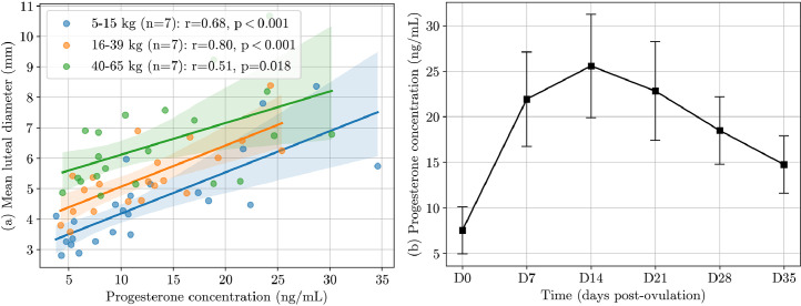
The corpus luteum is the only structure producing progesterone during pregnancy in dogs. The aim of this study was to characterise morphological changes of corpora lutea in the bitch and assess their relationship with body weight, serum progesterone concentration, and multiple resorptions. We monitored 26 bitches weekly from ovulation confirmation to 35 days post-ovulation, measuring the corpora lutea diameter via ultrasound examination in combination with progesterone assays. The pregnancy rate was 80.7% (21/26), and all pregnancies were carried to term. Dogs were classified into small (5-15 kg), medium (16-39 kg), and large breeds (40-65 kg). Dog weight significantly influenced mean luteal diameter (P < 0.001), which ranged from a mean ± SD of 3.4 ± 0.5 mm for small dogs to 6.0 ± 0.7 mm for large dogs on the day of ovulation confirmation. From ovulation confirmation to peak, corpora lutea grew significantly (2.1 ± 1.2 mm; P < 0.001) and returned to their initial size by day 35. Surprisingly, one-third of maximum corpora lutea exceeded 1 cm before undergoing subsequent physiological diametric reduction. This growth in luteal diameter was positively correlated with serum progesterone concentration (P < 0.05). This study provides novel findings on canine corpus luteum characteristics, not previously described in literature, which could aid ovulation detection and differentiation between physiological and potentially pathological structures.

Keywords: Bitch; Corpus luteum; Follicle; Pregnancy; Progesterone; Ultrasound.


Conflict of interest statement

The authors have no relevant financial or non-financial interests to disclose.

Figures

Fig 1
Fig. 1
Ovarian structures evolution in the same pregnant bitch (ID 18) during oestrus and dioestrus. Anechoic follicles (a: Day -3) show a reduction of their anechoic cavity, associated to peri-ovarian effusion (arrow, b: Day 0), at ovulation. The forming corpus luteum enlarges its anechoic lumen and thickens its wall (c: Day 8). Its development during dioestrus is characterised by a progressive antral wall thickening and obliteration of the central anechoic lumen (d: Day 21).
Fig 2
Fig. 2
Comparison of corpora lutea within the same ovary at different stages of wall thickening, observed at day 2 post-ovulation (left, ID 14) and day 14 post-ovulation (right, ID 11). The orange arrow indicates a nearly complete tissular corpus luteum, and the white arrow indicates a larger (1.5 cm) corpus luteum still presenting a large anechoic lumen. While large corpora lutea could be suspected of being ovarian cysts, the ones observed in this study gradually lost their anechoic lumen to become completely tissular by day 21.
Fig 3
Fig. 3
Boxplots showing maximum (a) and mean (b) luteal diameter (mm) evolution across days post-ovulation for 21 pregnant dogs. The box plot includes the median (line inside the box), interquartile range (box), outliers (dots), and mean (markers). Different superscript letters indicate statistical difference (P < 0.05) between days, as determined by an analysis of variance followed by Bonferroni's multiple comparison test.
Fig 4
Fig. 4
Scatter and bar plot of maximum (a) and mean (b) luteal diameters in 21 pregnant bitches measured weekly from day 0 to day 35 post-ovulation. Bitches are categorised into three weight groups, 5–15 kg (n = 7), 16–39 kg (n = 7), and 40–65 kg (n = 7). The bars represent the mean ± standard deviation across all days and the tables present the values for days 0, 7, 14, 21, 28, and 35 post-ovulation. Different superscript letters indicate statistical difference (P < 0.05) between body weight groups, as determined by an analysis of variance followed by Bonferroni's multiple comparison test.
Fig 5
Fig. 5
Left: Scatter plot showing the relationship between number of corpora lutea and body weight in 21 pregnant bitches. The regression line represents the linear fit, with the shaded area indicating the 95% confidence interval (Pearson's r = 0.86). Right: Bar plot of the number of corpora lutea categorised into three weight groups, 5–15 kg (n = 7), 16–39 kg (n = 7), and 40–65 kg (n = 7). Bars represent the mean ± standard deviation. Different superscript letters indicate statistical difference (P < 0.05) between weight groups, as determined by an analysis of variance.
Fig 6
Fig. 6
(a) Scatter plot, Pearson correlation coefficient, and P-value for progesterone concentration (ng/mL) versus mean luteal diameter in 21 pregnant bitches, among three body weight groups: 5–15 kg, 16–39 kg, 40–65 kg. Each point corresponds to an ovarian ultrasound performed concurrently with a blood sample between day 0 and 8 post-ovulation, and the continuous line with shaded regions represents the best-linear fit with 95% confidence intervals (CI). (b) Progesterone concentration (ng/mL) across days post-ovulation for 21 pregnant dogs, with mean values and standard deviations.

References

    1. Arashiro E.K.N., Viana J.H.M., Fonseca J.F.D., Camargo LSDA, Fernandes CADC, Brandão F.Z. Luteal dynamics in goats: morphological and endocrine features. Revista Brasileira de Zootecnia. 2010;39(9):1937–1942. doi: 10.1590/S1516-35982010000900011. - DOI
    1. Arlt S.P., Haimerl P. Cystic ovaries and ovarian neoplasia in the female dog - a systematic review. Reproduction in domestic animals = Zuchthygiene. 2016;51(Suppl 1):3–11. doi: 10.1111/rda.12781. - DOI - PubMed
    1. Bergeron L.H., Nykamp S.G., Brisson B.A., Madan P., Gartley C.J. An evaluation of B-mode and color Doppler ultrasonography for detecting periovulatory events in the bitch. Theriogenology. 2013;79(2):274–283. doi: 10.1016/j.theriogenology.2012.08.016. - DOI - PubMed
    1. Concannon P.W. Reproductive cycles of the domestic bitch. Animal reproduction science. 2011;124(3-4):200–210. doi: 10.1016/j.anireprosci.2010.08.028. - DOI - PubMed
    1. England G.C., Yeager A.E. Ultrasonographic appearance of the ovary and uterus of the bitch during oestrus, ovulation and early pregnancy. Journal of reproduction and fertility. 1993;47:107–117. - PubMed

LinkOut - more resources