

Genomic surveillance and evolutionary dynamics of respiratory syncytial virus circulating in Tunisia post-COVID-19 pandemic lockdown restrictions
- PMID: 40225290
- PMCID: PMC11987636
- DOI: 10.1016/j.ijregi.2025.100609
Genomic surveillance and evolutionary dynamics of respiratory syncytial virus circulating in Tunisia post-COVID-19 pandemic lockdown restrictions
Abstract
Objectives: Human respiratory syncytial virus (hRSV) is the leading cause of severe respiratory infections in children worldwide. Severe cases often require intensive care. Respiratory syncytial virus (RSV) is classified into two main antigenic groups: RSV-A and RSV-B. Recent molecular advancements have significantly enhanced our ability to control and understand RSV infections. This study investigated the epidemiologic and genetic characteristics of the RSV in Tunisia, focusing on its evolutionary dynamics.
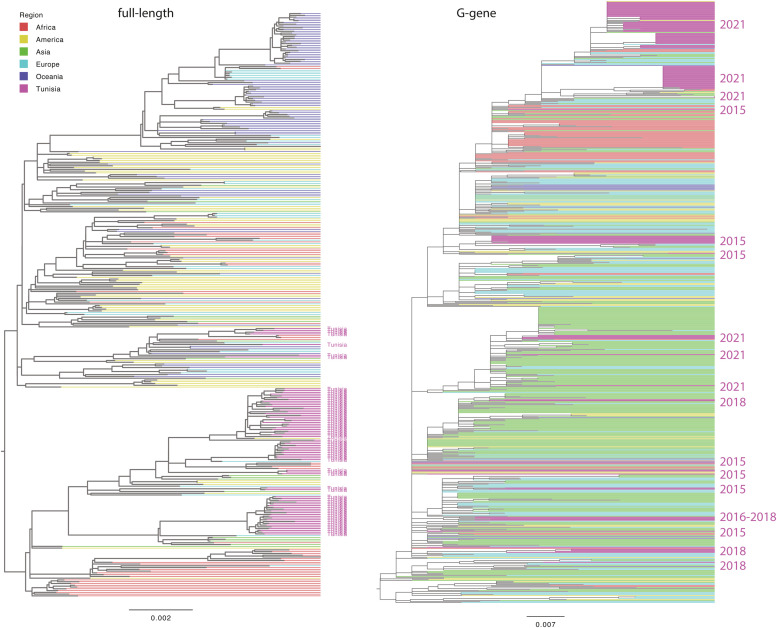
Methods: Between October and December 2021, 92 samples were collected from Tunisian patients hospitalized for mild-to-severe acute respiratory infections. Laboratory analyses, including real-time reverse transcription-polymerase chain reaction and whole genome sequencing (WGS), were performed to identify and characterize the RSV strains. Phylogenetic analyses were performed to compare the Tunisian sequences with the global RSV sequences from 2012 to 2022.
Results: Of the 92 patients (mean age 1 year and 6 months), 96.9% of the samples tested positive for RSV-A, and 22 samples exhibited co-infections with other respiratory viruses. WGS was successfully performed on the 74 samples. Phylogenetic analysis identified six distinct clades of RSV-A circulating in Tunisia, indicating multiple parallel introductions of the virus into the country. Specific Tunisian clades showed genetic similarities to RSV strains from Argentina, Belgium, and the Philippines.
Conclusions: This study underscores the genetic diversity of RSV in Tunisia, with multiple introductions after the lifting of COVID-19 restrictions in 2021. WGS revealed significant genetic heterogeneity within RSV-A, which could affect the effectiveness of vaccines and therapeutic strategies. Continued surveillance, particularly, in resource-limited regions, is crucial to inform and guide future interventions.
Keywords: Genetic diversity; Human respiratory syncytial virus (HRSV); Surveillance; Tunisia; Whole genome sequencing (WGS).
© 2025 The Author(s).
Conflict of interest statement
The authors declare no conflict of interest. The mention of trade names or commercial products in this article is solely for the purpose of providing specific information and does not imply a recommendation or endorsement by the IZSAM.
Figures


References
-
- Nair H., Nokes D.J., Gessner B.D., Dherani M., Madhi S.A., Singleton R.J., et al. Global burden of acute lower respiratory infections due to respiratory syncytial virus in young children: a systematic review and meta-analysis. Lancet. 2010;375:1545–1555. doi: 10.1016/S0140-6736(10)60206-1. - DOI - PMC - PubMed
LinkOut - more resources
Full Text Sources
