

High-Flow Congenital Arteriovenous Malformation of the Right Deltoid Muscle in a Pediatric Patient: A Case Report
- PMID: 40225515
- PMCID: PMC11994228
- DOI: 10.7759/cureus.80580
High-Flow Congenital Arteriovenous Malformation of the Right Deltoid Muscle in a Pediatric Patient: A Case Report
Abstract
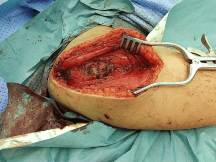
Congenital arteriovenous malformations (AVMs) are rare vascular anomalies involving abnormal connections between arteries and veins, bypassing the capillary bed. High-flow AVMs may lead to complications such as pain, ulceration, and functional impairment. We report a case of a six-year-old girl with a high-flow AVM in the right deltoid muscle, initially diagnosed via Doppler ultrasound and confirmed by MRI. The patient presented with a progressively enlarging, painless swelling of the right shoulder. Management included super-selective embolization, followed by surgical resection due to symptom recurrence and persistent vascular shunting. Postoperatively, the patient experienced improved shoulder mobility and pain reduction. The treatment plan involved a staged approach, with initial embolization to reduce blood flow, followed by surgical excision to prevent recurrence. The surgical resection was performed soon after the second embolization. Postoperative care included pain management and physiotherapy for optimal recovery. This case emphasizes the importance of early diagnosis, multimodal intervention, and long-term follow-up in pediatric AVMs. Future studies should focus on recurrence predictors, optimal timing for surgical resection post-embolization, and the role of genetic factors in AVM development.
Keywords: arteriovenous malformations; embolization; pediatric; surgical resection; vascular anomalies.
Copyright © 2025, Zaid et al.
Conflict of interest statement
Human subjects: Consent for treatment and open access publication was obtained or waived by all participants in this study. Conflicts of interest: In compliance with the ICMJE uniform disclosure form, all authors declare the following: Payment/services info: All authors have declared that no financial support was received from any organization for the submitted work. Financial relationships: All authors have declared that they have no financial relationships at present or within the previous three years with any organizations that might have an interest in the submitted work. Other relationships: All authors have declared that there are no other relationships or activities that could appear to have influenced the submitted work.
Figures


Similar articles
-
Surgical resection of a rare scalp arteriovenous malformation: a case report.Ann Med Surg (Lond). 2024 Sep 24;86(11):6805-6809. doi: 10.1097/MS9.0000000000002596. eCollection 2024 Nov. Ann Med Surg (Lond). 2024. PMID: 39525744 Free PMC article.
-
Successful Management of a Rare Case of Arteriovenous Malformation of the Forearm in a 15-Year-Old Girl: A Case Report.J Orthop Case Rep. 2025 Apr;15(4):132-135. doi: 10.13107/jocr.2025.v15.i04.5473. J Orthop Case Rep. 2025. PMID: 40212479 Free PMC article.
-
Arteriovenous Malformation of the Upper Eyelid: A Case Report.Plast Reconstr Surg Glob Open. 2021 Jun 10;9(6):e3609. doi: 10.1097/GOX.0000000000003609. eCollection 2021 Jun. Plast Reconstr Surg Glob Open. 2021. PMID: 34123685 Free PMC article.
-
Management of extracranial arteriovenous malformations of the head and neck.Auris Nasus Larynx. 2020 Apr;47(2):181-190. doi: 10.1016/j.anl.2019.11.008. Epub 2019 Dec 18. Auris Nasus Larynx. 2020. PMID: 31862283 Review.
-
Consensus Document of the International Union of Angiology (IUA)-2013. Current concept on the management of arterio-venous management.Int Angiol. 2013 Feb;32(1):9-36. Int Angiol. 2013. PMID: 23435389
References
-
- Differentiation of vascular birthmarks by MR imaging. An investigation of hemangiomas, venous and lymphatic malformations. Kern S, Niemeyer C, Darge K, Merz C, Laubenberger J, Uhl M. Acta Radiol. 2000;41:453–457. - PubMed
-
- Arteriovenous malformations of the temporalis muscle: a comprehensive review. Ros de San Pedro J, Cuartero Pérez B, Ferri Ñíguez B, Villanueva San Vicente V. Oper Neurosurg (Hagerstown) 2018;14:325–340. - PubMed
-
- Novel multimodality imaging techniques for diagnosis and evaluation of arteriovenous malformations. Mokin M, Dumont TM, Levy EI. Neurol Clin. 2014;32:225–236. - PubMed
Publication types
LinkOut - more resources
Full Text Sources