

Clinical Features and Imaging Findings of Low-Grade Endometrial Stromal Sarcoma: A Retrospective Case Series-Based Analysis
- PMID: 40225539
- PMCID: PMC11993436
- DOI: 10.7759/cureus.80507
Clinical Features and Imaging Findings of Low-Grade Endometrial Stromal Sarcoma: A Retrospective Case Series-Based Analysis
Abstract
Objective: This case series presents six cases of low-grade endometrial stromal sarcoma (LGESS) and discusses their magnetic resonance imaging (MRI) findings in light of recent advancements in LGESS diagnosis.
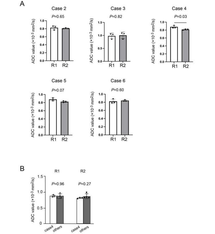
Methods: Data from six cases of LGESS treated at Fukui University Hospital between June 2007 and October 2024 were retrospectively analyzed. The clinical behavior, histopathological features, therapeutic approaches, and survival outcomes were evaluated. MRI images were assessed by two radiologists over multiple parameters that were considered specific to the LGESS.
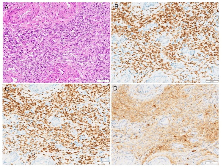
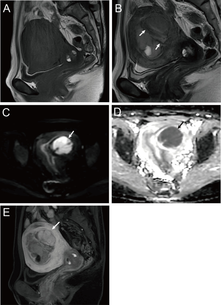
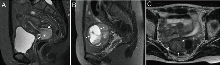
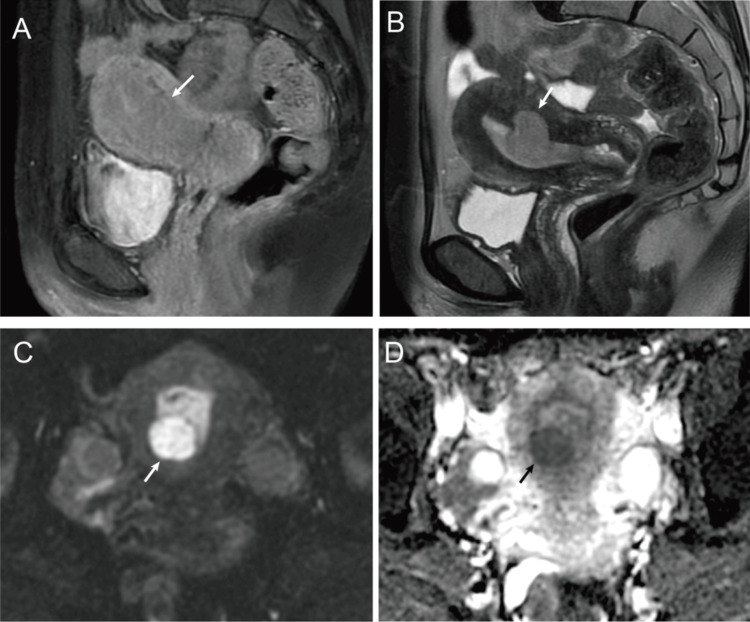
Results: Among the six patients with LGESS, four patients had tumors located within the myometrium, whereas two patients had tumors located in the submucosal layer. All cases were classified as International Federation of Gynecology and Obstetrics Stage Ⅰ, and no recurrence was observed during follow-up. On MRI, all six tumors exhibited high signal intensity on T2-weighted images (T2WI), compared to the myometrium, and high signal intensity on diffusion-weighted images (DWI), compared to the endometrium. In four of the five evaluable cases, the mean apparent diffusion coefficient (ADC) value was 0.86 × 10⁻³ mm²/s (range: 0.81-0.99).
Conclusion: Both clinically and based on imaging findings, distinguishing between LGESS and rare leiomyoma variants is challenging. MRI findings, including high signal intensity on T2WI and DWI as well as low ADC values, may prove valuable in differentiating these two entities.
Keywords: apparent diffusion coefficient (adc); diffusion-weighted imaging (dwi); endometrial stromal sarcoma; leiomyoma uteri; magnetic resonance imaging.
Copyright © 2025, Miyazaki et al.
Conflict of interest statement
Human subjects: Consent for treatment and open access publication was obtained or waived by all participants in this study. Animal subjects: All authors have confirmed that this study did not involve animal subjects or tissue. Conflicts of interest: In compliance with the ICMJE uniform disclosure form, all authors declare the following: Payment/services info: All authors have declared that no financial support was received from any organization for the submitted work. Financial relationships: All authors have declared that they have no financial relationships at present or within the previous three years with any organizations that might have an interest in the submitted work. Other relationships: All authors have declared that there are no other relationships or activities that could appear to have influenced the submitted work.
Figures
Similar articles
-
Low-Grade Endometrial Stromal Sarcoma: A Case Report of a Rare Uterine Malignancy Mimicking Degenerative Uterine Leiomyoma in a Nulliparous Woman.Diagnostics (Basel). 2024 Dec 25;15(1):18. doi: 10.3390/diagnostics15010018. Diagnostics (Basel). 2024. PMID: 39795543 Free PMC article.
-
Differentiation of uterine low-grade endometrial stromal sarcoma from rare leiomyoma variants by magnetic resonance imaging.Sci Rep. 2021 Sep 27;11(1):19124. doi: 10.1038/s41598-021-98473-z. Sci Rep. 2021. PMID: 34580348 Free PMC article.
-
Focal Area of Low T2 Signal on MRI Scans in a Heterogeneous Uterine Leiomyoma Does Not Exclude the Possibility of Malignancy: A Report of Two Cases.Case Rep Radiol. 2025 Apr 25;2025:5388015. doi: 10.1155/crra/5388015. eCollection 2025. Case Rep Radiol. 2025. PMID: 40322071 Free PMC article.
-
Uterine Endometrial Stromal Tumors With Pure Low-Grade Morphology Harboring YWHAE::NUTM2 Fusions: Report of a Case Series Emphasizing Potential for High-Grade Transformation and Aggressive Behavior.Am J Surg Pathol. 2023 Jun 1;47(6):717-724. doi: 10.1097/PAS.0000000000002041. Epub 2023 Apr 10. Am J Surg Pathol. 2023. PMID: 37032555
-
A Rare Case of Low-Grade Endometrial Stromal Sarcoma Invading an Old Leiomyoma.Cureus. 2024 Nov 9;16(11):e73329. doi: 10.7759/cureus.73329. eCollection 2024 Nov. Cureus. 2024. PMID: 39655119 Free PMC article.
References
-
- Low-grade endometrial stromal sarcoma-a review. Thiel FC, Halmen S. Oncol Res Treat. 2018;41:687–692. - PubMed
-
- Endometrial stromal tumors of the uterus: epidemiology, pathological and biological features, treatment options and clinical outcomes. Gadducci A, Multinu F, De Vitis LA, Cosio S, Carinelli S, Aletti GD. Gynecol Oncol. 2023;171:95–105. - PubMed
LinkOut - more resources
Full Text Sources