Review
. 2025 Mar 31;15(10):4823-4847.
doi: 10.7150/thno.108875. eCollection 2025.

Macrophage membrane-functionalized nanotherapeutics for tumor targeted therapy

Affiliations
Review

Macrophage membrane-functionalized nanotherapeutics for tumor targeted therapy

Mubassir Khan et al. Theranostics. .

Abstract

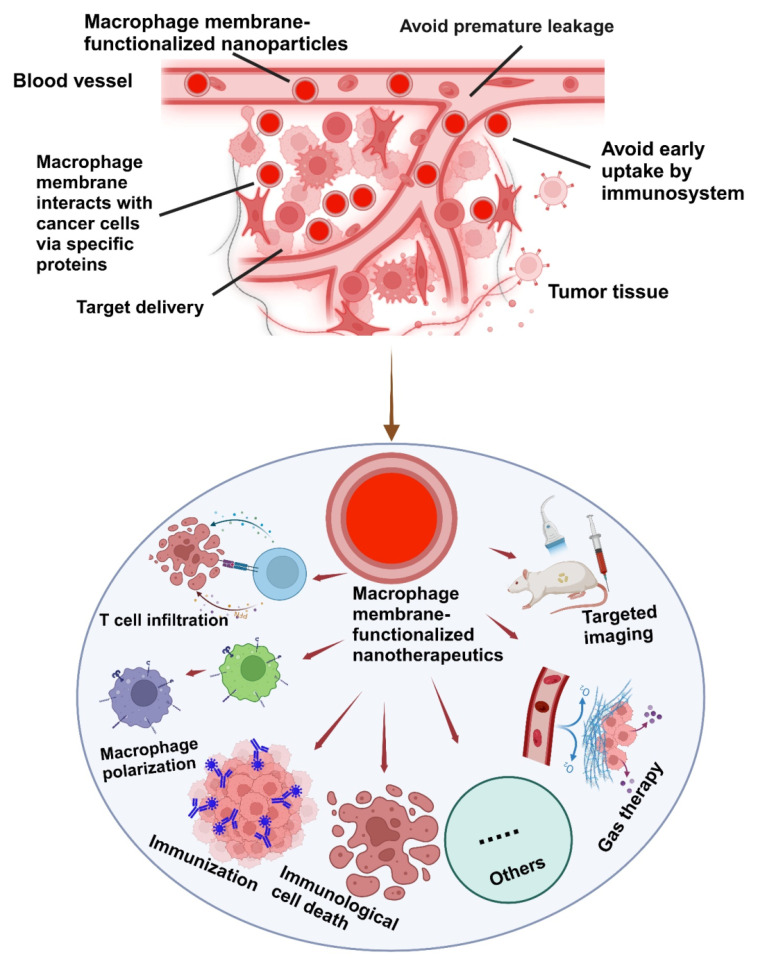
Cancer is a multifaceted disease characterized by uncontrollable cell growth. To date, various therapies are employed including conventional chemotherapy, surgery, radiotherapy, and immunotherapies. However, these approaches still present significant limitations. Interestingly, macrophage membranes utilize their innate antigen recognition affinity to facilitate targeted localization to tumor sites with high specificity. As a result, they display distinct characteristics such as avoiding premature leakage, tumor targeting ability, immune evasion, immune system activation, tumor-infiltrating ability, improved cell endocytosis and release payload in tumor-microenvironment. In this paper, the recent advances in macrophage-membrane-encapsulated nanotherapeutics for targeted cancer therapy are presented. We begin by introducing macrophage membrane-encapsulated nanotherapeutics preparation and characterization, followed by cancer immunotherapy such as macrophage polarization, T-cell infiltration, macrophage membrane modification, immunization, and inducing immunological cell death. Lastly, a future perspective is proposed to highlight the limitations of macrophage membrane-encapsulated nanotherapeutics and the possible resolutions toward the clinical transformation of currently developed biomimetic chemotherapies. We believe this review may be beneficial for improving the deep research of macrophage membrane-encapsulated nanotherapeutics for targeted cancer therapy.

Keywords: cancer therapy; immunological cell death; macrophage membrane; nanotherapeutics; targeted delivery.


Conflict of interest statement

Competing Interests: The authors have declared that no competing interest exists.

Figures

Figure 1
Figure 1
This figure highlights the development of natural biomembrane-based nanotherapeutics for tumor-targeted therapy during the past decade. It demonstrates the use of different cell membranes as drug carriers, emphasizing their potential to improve targeted drug delivery.
Figure 2
Figure 2
Schematic illustration of the possible mechanisms of MΦM-coated nanotherapeutics exhibiting high tumor-targeted delivery efficiency. Their inherent membrane characteristics enable effective tumor targeting, evasion of the immune system, avoidance of premature leakage, and enhanced cellular absorption improving therapeutic effectiveness while reducing off-target effects and systemic toxicity in tumor-targeted therapy.
Figure 3
Figure 3
Preparation methods for MΦM. (A) Schematic diagram of the preparation of atorvastatin and polydatin co-loaded MΦM-coated metal-organic framework NPs (AP@ZIF-Mem). Reproduces with permission . Copyright 2024, Elsevier. (B) Extrusion method to create FBN@M. Reproduce with permission . Copyright, 2024 American Chemical Society. (C) Synthesis strategy through the combination of MNC synthesis, an engineered membrane with an azide group, electrostatic assembly, and click reaction. Reproduced with permission . Copyright 2018, Wiley Online Library.
Figure 4
Figure 4
MΦM-coated NP characterization. (A) NPR@TAMMs images (a) TEM analysis of NPR@TAMMs. (b) The up-conversion emission spectrum of UCNPs and NPR@TAMMs. (c) Western blotting for quantification of different cell membrane markers for NPR, cell lysate, TAMMs, and NPR@TAMMs. (d) Hydrodynamic size of NPR@TAMMs. (e) Generation of singlet O2 by NPR@TAMMs based on the fluorescence intensity. (f) Zeta-potential of NPR@TAMMs. Reproduced with permission . Copyright 2021, American Chemical Society. (B) (a) TEM analysis of UCNPs@mSiO2-PFC/Ce6, UCNPs@mSiO2-PFC/Ce6@RAW, UCNPs@mSiO2-PFC/Ce6@RAW-Man, and UCNPs@mSiO2-PFC/Ce6@RAW-Man/PTX. (b) Magnified TEM images of UCNPs@mSiO2-PFC/Ce6 and UCNPs@mSiO2-PFC/Ce6@RAW-Man/PTX and their element mappings for Na, F, Er, Lu, Si, and P. Reproduced with permission . Copyright 2023, Elsevier. (C) RAW M@MBG (c1-6). Reproduced with permission . Copyright 2023, Elsevier (D) TEM image of FBN@M and SDS-PAGE protein analysis of MΦM on FBN@M. Reproduced with permission . Copyright 2024, American Chemical Society. (E) M@C-HA/ICG SEM analysis of M, and M@C-HA/ICG. Reproduced with permission . Copyright 2022, Elsevier.
Figure 5
Figure 5
Targeting tumors and polarizing M2 tumor-associated macrophages to M1 phenotype (A) CLSM images of M2 RAW 264.7 cells after treatment with DiI-stained UC-NPs. (B) Relative expression of M1-markers [(a) CD86, (b) TNF-α, and (c) iNOS] and M2 markers (d) CD206 and (e) TGF-β as shown by qRT-PCR: Treatment A = Ce6 + PTX + 808 nm; B = UCNPs@mSiO2-PFC/Ce6 + 808 nm; C = UCNPs@mSiO2-PFC/Ce6@RAW-Man/PTX + 808 nm. (f) Relative expression levels of M1 and M2 markers. (C) CLSM images of RAW 264.7 cells treated with PBS, LPS/IFN-γ, and IL-4 in normoxic conditions showing CD86/CD206 expression. (D) Flow cytometry analysis of RAW 264.7 cells for M1, M2, and M2 + Treat =C. (E) Representative image of SDS-gel of M1 and M2 types (a) and western blot for cell membrane markers for M0, M1, M2, and M2 + Treat=C (b). Reproduced with permission . Copyright 2023, Elsevier. (B) Immunological regulations of M1/PLGA@IR780/CAT. Representative immunofluorescent staining images: red of M1-marker, CD86 (A) and M2-marker CD206 (C), and blue (DAPI). (B) The relative quantification of CD86, (D), and CD206 after different concentration treatments. (E, G) Flow cytometry analysis of the population of M1/M2-macrophage after different concentration treatments. (F, H) The percentages of M1-macrophage (F4/80 + CD86+) and M2-macrophage (F4/80+ CD206+). (I) Secretion of TNF-a, IL-10, and IL-6 after different concentration treatments. Reproduced with permission . Copyright 2024, Elsevier. (C) M@C-HA/ICG and macrophage polarization. (a, b) The ratio of M1- macrophages after being treated with LPS and C-HA/ICG. (c) The generation of ROS, NO, TNF-α, and IL-12 after RAW264.7 cells were treated with different concentrations. (d) The release of LDH from 4T1 cells after treatment with M or M@C-HA/ICG. (e) Fluorescent images of 4T1 cells labeled with calcein-AM/PI after co-culture with M or M@C-HA/ICG. (f) Toxicity of M@C-HA/ICG to 4T1 cells. Reproduced with permission . Copyright 2022, Elsevier. (D) In vivo macrophage polarization for a pro-inflammatory TME in tumor mice. (A) The mRNA levels of M1 (iNOS and TNF-α) and M2 markers (Arg-1 and IL-10) within the tumor tissues using qRT-PCR. (B) TAM repolarization: representative immunofluorescence images of iNOS (red) and Arg-1 (red) from confocal microscopy. Reproduced with permission copyright 2022, American Chemical Society.
Figure 6
Figure 6
Immunomodulatory effects of MΦM-functionalized PCL nanofibers. The PCL nanofibers are wrapped with MΦM from different macrophages M0, M1, and M2 phenotypes. Various types of influences of the two types of nanofibers are presented for a direct comparison. Reproduced with permission . Copyright 2022, Elsevier.
Figure 7
Figure 7
T-Cell Infiltrations (A) Schematic Illustration of the Drug-Loaded Hydrogel (Gel@M/CuO2/DOX/STING) for Preventing Postoperative Tumor Recurrence and Metastasis. Reproduced with permission . Copyright 2023, American Chemical Society. (B) M1HD@RPR preparations and its targeted mechanism in combined cancer therapy. Reproduced with permission , copyright 2022, science. (C) Schematic diagram of the synthesis of RAW 264.7-PD-1 and PD-1-MM@PLGA/RAPA and engineered MΦM-coated-NPs with enhanced PD-1 expression. Reproduced with permission . Copyright 2022, American Chemical Society.
Figure 8
Figure 8
PCoA@M preparations PDA, ADT, and Co-MOF were prepared by a one-pot method and modified with MΦM to form PCoA@M and enriched in the TME via targeting integrin. PCoA@M enhanced PTT by blocking HSPs, degrading and releasing drugs under the TME acidic conditions, reducing NADH generation, and achieving PTT gas synergistic starvation therapy. Reproduced with permission . Copyright 2022, Nature.
Figure 9
Figure 9
Schematic illustration showing the integration of NIR-AIEgen with mesoporous PB nano-catalyzer boosts the theranostic performance for NIR-II fluorescence and PA imaging-guided robust cancer immunotherapy. Reproduced with permission , copyright 2024, Wiley online library.
Figure 10
Figure 10
The illustration demonstrates that the hypoxia-activated nanoplatform integrates NIR-II fluorescence (NIR-II FL) and photoacoustic (PA) imaging for precise tumor localization, along with synergistic immunotherapy via TME remodeling, ICD, and STING pathway activation to enhance cancer treatment. Reproduced with permission , copyright 2024, Nature.
Figure 11
Figure 11
Macrophage membrane conjugated biomolecules and immunization (A) Schematic illustration of the preparation of ARMFU. M1-macrophages engineered with ARMFUL and CSF1R-inhibitor BLZ945 were loaded in the PLGA-based polymeric core, and aCD47 was conjugated on the fusogenic lipid shell surface, showing in the TEM image a aCD47, BLZ@MFUL, and ARMFUL. This ARMFUL can fuse with the M1-MΦM to simultaneously insert aCD47-modified lipid shells on the surfaces directly and release the BLZ945-loaded core into the cytoplasm, formulating ARMFUL/M1 for back-transfer and could remodel the tumor microenvironment. ARMFUL/M1 macrophages could remodel the TME, activate T-cell cytotoxicity, and induce systemic immunological memory to synergistically inhibit tumor growth. Reproduced with permission . Copyright 2023, science. (B) Illustration of antiHER2-engineered macrophage biomimetic photothermal (AMBP) systems for photothermal/biotherapy of cancer. Reproduced with permission . Copyright 2024, Elsevier.

Similar articles

Cited by

References

    1. Hanahan D. Hallmarks of cancer: new dimensions. Cancer Discov. 2022;12(1):31–46. - PubMed
    1. Kratz CP, Jongmans MC, Cavé H, Wimmer K, Behjati S, Guerrini-Rousseau L. et al. Predisposition to cancer in children and adolescents. Lancet Child Adolesc Health. 2021;5(2):142–54. - PubMed
    1. Rivlin N, Brosh R, Oren M, Rotter V. Mutations in the p53 tumor suppressor gene: important milestones at the various steps of tumorigenesis. Genes Cancer. 2011;2(4):466–74. - PMC - PubMed
    1. Chung SW, Kim GC, Kweon S, Lee H, Choi JU, Mahmud F. et al. Metronomic oral doxorubicin in combination of Chk1 inhibitor MK-8776 for p53-deficient breast cancer treatment. J Biomater. 2018;182:35–43. - PubMed
    1. Bowerman CJ, Byrne JD, Chu KS, Schorzman AN, Keeler AW, Sherwood CA. et al. Docetaxel-loaded PLGA nanoparticles improve efficacy in taxane-resistant triple-negative breast cancer. Nano Lett. 2017;17(1):242–8. - PMC - PubMed

MeSH terms

Substances