. 2025 Mar 28:16:1547649.
doi: 10.3389/fmicb.2025.1547649. eCollection 2025.

Rhomboid proteases: key players at the cell surface within haloarchaea

Affiliations

Rhomboid proteases: key players at the cell surface within haloarchaea

Mariana Inés Costa et al. Front Microbiol. .

Abstract

Introduction: Rhomboid proteases are intramembrane serine proteases that play a key role in regulating membrane proteins across all domains of life. However, their function in archaea remains poorly understood. The model halophilic archaeon Haloferax volcanii encodes two rhomboid homologs, rho1 (HVO_1474) and rho2 (HVO_0727). Previous studies indicated that the deletion of rho2 resulted in mild alterations in motility, adhesion, biofilm formation, and cell morphology, suggesting potential functional compensation by rho1.

Materials and methods: To investigate the role of these proteases, we generated single (Δrho1) and double (Δrho1 Δrho2) deletion mutants. Phenotypic characterization included viability assays, motility tests, adhesion and biofilm formation studies, as well as morphological analysis using microscopy. Functional overlap between rho1 and rho2 was evaluated through genetic complementation/overexpression experiments in which each gene was expressed in trans in the mutant backgrounds.

Results: Both Δrho1 and Δrho1 Δrho2 mutants were viable, indicating that these genes are not essential in H. volcanii. The Δrho1 mutant exhibited increased motility, enhanced biofilm formation, reduced adhesion to glass surfaces, and significant morphological alterations, particularly in trace element-deficient conditions. The double mutant (Δrho1 Δrho2) showed increased adhesion to surfaces, mild motility reduction, and fewer morphological abnormalities compared to Δrho1. Complementation assays revealed that both rho1 and rho2 could restore motility in Δrho2 and adhesion in Δrho1. However, only rho1 was able to complement the morphological defects, suggesting a degree of functional divergence between these homologs.

Discussion: This work highlights the role of rhomboid proteases in regulating critical cell surface processes in H. volcanii, including biofilm formation, surface adhesion, and cell shape determination. The ability of rho1 and rho2 to compensate for each other in certain functions while maintaining distinct roles underscores a complex regulatory interplay. Future research will focus on identifying natural substrates and elucidating the molecular mechanisms underlying rhomboid protease function in haloarchaea.

Keywords: Haloferax volcanii; archaeal morphology; haloarchaea; intramembrane proteases; rhomboid protease.


Conflict of interest statement

The authors declare that the research was conducted in the absence of any commercial or financial relationships that could be construed as a potential conflict of interest.

Figures

Figure 1
Figure 1
Complementation of the motility phenotype in the Δrho2 mutant with plasmids expressing rho homologs. H. volcanii H26 pTA963 and Δrho2 transformed with either pTA963, pTA963::rho1, pTA963::rho2, or pMCF1 (containing the complete rho2/endV operon, under the endogenous promoter) were cultured in CAB medium with 2 mM trp at 42°C overnight (ON) and subsequently adjusted to an OD600 = 0.5. For the motility assay, 1 μL of the adjusted culture was inoculated onto swimming agar plates with 2 mM trp and incubated for 48 h. Photographs were taken and the ring diameter was measured using ImageJ. All strains were inoculated on the same plate and three independent experiments with five replicates each were conducted. The results were analyzed using the R program. (A) Box plot of the distribution of the motility rings diameters for each strain. (B) Representative image of a soft agar motility plate.
Figure 2
Figure 2
Evaluation of Rho deletion mutants growth in liquid medium. Cultures of the indicated strains were inoculated from ON starter cultures at an initial OD600 of 0.01 in 3 mL of CAB medium supplemented with ura (50 μg/mL). Growth was monitored by measuring OD600 at the indicated times and the values were used to generate the growth curve (A) Each culture was grown as three biological replicates, with experiments independently repeated at least three times. Growth rates were determined during the E phase by fitting linear regression models to the OD600 data and calculating the slopes, using GraphPad Prism software. For comparison, the H26, Δrho1, Δrho2 and Δrho1 Δrho2 strains growth rate was plotted against the H26 growth curve, as a reference growth curve (B) Statistical significance was assessed using a two-tailed Student’s t-test (p-value <0.05 considered significant).
Figure 3
Figure 3
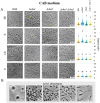
Cell morphology of H. volcanii Rho mutants in CA medium. H. volcanii single colonies of the specified strains were inoculated into 3 mL cultures of CA medium and samples were taken at EE (OD600 0.1–0.3), E (OD600 0.4–0.6), LE (OD600 0.7–1), and S phases (OD600 ≥ 1.3). (A) Cells were imaged in an inverted microscope under 0.5% (w/v) CAB-agarose pads. Right panel shows the distribution of the cell’s aspect ratio for each strain and growth phase. At least 10 photograms from three independent cultures were analyzed for each strain and condition. (B) Different morphological abnormalities observed for the Δrho1 strain in CA medium.
Figure 4
Figure 4
Cell morphology of H. volcanii Rho mutants in CAB medium. H. volcanii single colonies of the specified strains were inoculated into 3 mL cultures of CAB and samples were taken at the same stages of the growth curve depicted in Figure 3. (A) Cells were imaged in an inverted microscope under 0.5% (w/v) CAB-agarose pads. Right panel shows the distribution of cell aspect ratio for each strain and growth phase. At least 10 photograms from three independent cultures were analized for each strain and condition. (B) Morphological abnormalities observed for the Δrho1 strain in CAB medium.
Figure 5
Figure 5
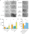
Complementation of the abnormal morphology phenotype in the Δrho1 strain. H. volcanii H26 and Δrho1 strains were transformed with either pTA963, pTA963::rho1 or pTA963::rho2 plasmids and cultured in CAB (A) or CA (B) medium until reaching the EE growth phase. Growth was conducted in the presence or absence of 2 mM trp to induce gene expression, as indicated on the left. Cell morphology was observed using optical microscopy as described in the Materials and Methods section. The photograms are representative of three independent cultures for each condition tested.
Figure 6
Figure 6
Effect of Rho gene elimination on cell motility in H. volcanii. Single colonies of the indicated strains were stab-inoculated on CAB soft agar plates containing 50 μg/mL ura and incubated at 42°C for 48–72 h. After incubation, photographs were taken, and the diameters of the growth halos were measured using ImageJ software. (A) Diameter values corresponding to three independent experiments, each with five replicates. Results were analyzed by LMMs (linear mixed models) with normal distribution to determine statistical significance between treatment and controls. A post hoc analysis was conducted using Tukey. All analyses were performed using R software version 3.6.1 (R Core Team, 2019). Statistically significant differences were determined at p < 0.05. (B) Representative photograph showing the motility halos generated by each strain.
Figure 7
Figure 7
Evaluation of cell adhesion in Rho mutants. Glass coverslips were immersed in triplicate cultures of H. volcanii strains and incubated at 42°C for 1 or 3 h, as specified, followed by staining with crystal violet as described in the Materials and Methods section. (A,C) Optical microscopy images (1000×) showing cell adhesion to the glass surface for each strain. (B,D) Quantification of three independent experiments using Image J software. Data were analyzed using linear models (LM) with random effects, followed by Tukey’s post hoc test. Statistical analyses were performed with R software version 3.6.1 (R Core Team, 2019). Differences were considered statistically significant at p < 0.05.
Figure 8
Figure 8
Effect of Rho gene deletion on biofilm formation in H. volcanii. EE phase cultures were transferred to 96-well polystyrene plates and incubated without agitation at 42°C for 72 h. Early-stage biofilms were stained with crystal violet according to the Materials and Methods section. Data was obtained from three independent experiments, each including six technical replicates. The amount of cell-bound colorant was quantified by measuring A574, and values corresponding to the mutant strains were normalized to the average value of the parental strain (H26). Results were analyzed by LMMs (linear mixed models) with normal distribution to determine statistical significance between treatment and controls. A post hoc analysis was conducted using Tukey. All analyses were performed using R software version 3.6.1 (R Core Team, 2019). Statistically significant differences were determined at p < 0.05.

Similar articles

References

    1. Abdul-Halim M. F., Karch K. R., Zhou Y., Haft D. H., Garcia B. A., Pohlschroder M. (2015). Permuting the PGF signature motif blocks both Archaeosortase-dependent C-terminal cleavage and Prenyl lipid attachment for the Haloferax volcanii S-layer glycoprotein. J. Bacteriol. 198, 808–815. doi: 10.1128/JB.00849-15, PMID: - DOI - PMC - PubMed
    1. Allers T. (2009). The Halohandbook: Protocols for haloarchaeal genetics. Melbourne, VI, Australia: Haloarchaeal Genetics Laboratory.
    1. Allers T., Barak S., Liddell S., Wardell K., Mevarech M. (2010). Improved strains and plasmid vectors for conditional overexpression of his-tagged proteins in Haloferax volcanii. Appl. Environ. Microbiol. 76, 1759–1769. doi: 10.1128/AEM.02670-09, PMID: - DOI - PMC - PubMed
    1. Allers T., Ngo H. P., Mevarech M., Lloyd R. G. (2004). Development of additional selectable markers for the halophilic archaeon Haloferax volcanii based on the leuB and trpA genes. Appl. Environ. Microbiol. 70, 943–953. doi: 10.1128/AEM.70.2.943-953.2004, PMID: - DOI - PMC - PubMed
    1. Baker R. P., Wijetilaka R., Urban S. (2006). Two plasmodium rhomboid proteases preferentially cleave different adhesins implicated in all invasive stages of malaria. PLoS Pathog. 2:e113. doi: 10.1371/journal.ppat.0020113, PMID: - DOI - PMC - PubMed

LinkOut - more resources