. 2023 Aug 29:2023:6658543.
doi: 10.1155/2023/6658543. eCollection 2023.

Tendon Extracellular Matrix Promotes Myotendinous Junction Protein Expression in Engineered Muscle Tissue under Both Static and Mechanically Stimulated Culture Conditions

Affiliations

Tendon Extracellular Matrix Promotes Myotendinous Junction Protein Expression in Engineered Muscle Tissue under Both Static and Mechanically Stimulated Culture Conditions

Lewis S Gaffney et al. J Tissue Eng Regen Med. .

Abstract

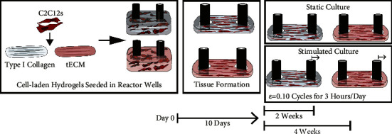
Studying the crosstalk between the muscle and tendon tissue is an important yet understudied area in musculoskeletal research. In vitro models can help elucidate the function and repair of the myotendinous junction (MTJ) under static and dynamic culture conditions using engineered muscle tissues. The goal of this study was to culture engineered muscle tissues in a novel bioreactor in both static and mechanically stimulated cultures and evaluate the expression of MTJ-specific proteins within the muscle-tendon unit(paxillin and type XXII collagen). C2C12 myoblasts were seeded in hydrogels made from type I collagen ortendon-derived extracellular matrix (tECM) and allowed to form around movable anchors. Engineered tissues were allowed to form and stabilize for 10 days. After 10 days in the culture, stimulated cultures were cyclically stimulated for 3 hours per day for 2 and 4 weeks alongside static cultures. Strain values at the maximum displacement of the anchors averaged about 0.10, a target that has been shown to induce myogenic phenotype in C2C12s. Protein expression of paxillin after 2 weeks did not differ between hydrogel materials in static cultures but increased by 62% in tECM when mechanically stimulated. These differences continued after 4 weeks, with 31% and 57% increases in tECM tissues relative to type I collagen. Expression of type XXII collagen was similarly influenced by hydrogel material and culture conditions. Overall, this research combined a relevant microenvironment to study muscle and tendon interactions with a novel bioreactor to apply mechanical strain, an important regulator of the formation and maintenance of the native MTJ.


Conflict of interest statement

The authors declare no conflicts of interest.

Figures

Figure 1
Figure 1
Loading of engineered tissues within the bioreactor. (a) Custom bioreactors were 3D printed with high-temperature polylactic acid. (b) Cell-laden hydrogels were seeded into custom inserts; after cells contract the hydrogels, tissues form around the anchor posts in the reactor. (c) To apply cyclic strain, the well plate is secured to a Velmex BiSlide® linear actuator with a stepper motor and a VXM controller. (d) During loading, the reactor system is placed in the incubator with a cooling plate connected to a radiator to reduce the temperature of the stepper motor by constantly circulating room-temperature fluid through the plate.
Figure 2
Figure 2
Experimental methods to study the effects of mechanically stimulating C2C12 and ECM hydrogel tissue constructs. Briefly, C2C12 myoblast cells were seeded in either type I collagen or tECM hydrogels; then, the cell-laden hydrogel was transferred to a reactor well. Within the culture wells, the cells contracted the hydrogel-forming tissue around the reactor posts after 10 days. Tissues were either cultured in a static condition with no cyclic strain loading or loaded to a strain of 0.10 cyclically for 3 hr per day. Tissues were cultured for 2 and 4 weeks before endpoint analysis to study gene and protein expression.
Figure 3
Figure 3
To verify the calculated displacement resulted in the expected strain, tissues were analyzed with DIC to determine strain across 3 days of mechanical stimulation. (a) Over three days, tissues were observed for 5 min before and after 3 hr of mechanical stimulation. (b) Videos acquired were rendered into individual images for DIC, and the region of interest was defined in Photoshop by masking individual tissues without the posts. (c) After DIC analysis, tissues had uniform strain throughout at peak and minimum displacement around the target values. (d) After tissue formation (day 10) and before any 3-hour mechanical stimulation, 10 loading cycles resulted in strains averaging 0.10 at maximum displacement and 0.01 at minimum displacement. (e) Over 3 days of observation, before and after 3 hours of loading, strains at maximum displacement averaged 0.10 and 0.00 at minimum displacement.
Figure 4
Figure 4
Relative gene expression determined with qPCR of Pax and Col22a1. (a) Expression of Pax was not significantly different in different hydrogel materials in static and stimulated cultures. (b) Col22a1 gene expression was markedly upregulated in tECM hydrogels compared to type I collagen (∗∗∗∗indicates p < 0.0001; indicates p < 0.05).
Figure 5
Figure 5
Paxillin protein expression was more evident in tECM tissues and highest in tECM tissues stimulated during the culture. (a, b) Representative images of samples stained with antipaxillin (red) and DAPI (blue) at 2 and 4 weeks, and scale bars are 200 μm. At both time points, staining is more evident in tECM tissues, specifically those with loading. (c, d) E.I. values were calculated by normalizing the positive area of the antipaxillin fluorescent channel, TXRED, and the DAPI fluorescent channel. Student's t-tests were used to compare the hydrogel materials in static or stimulated conditions. Large differences in paxillin expression are not seen after 2 weeks of the static culture; in the stimulated culture conditions, there was a significant difference between hydrogel materials (∗∗indicates p < 0.01). After 4 weeks, statistical differences between materials in both the culture conditions were observed.
Figure 6
Figure 6
Type XXII collagen expression was more evident in tECM tissues and highest in tECM tissues stimulated during the culture. (a, b) Representative images of samples stained with anti-Col22a1 (red) and DAPI (blue) at 2 and 4 weeks, and scale bars are 200 um. In each, staining is more evident in tECM tissues and is more prevalent throughout the tissues in stimulated tECM tissues. (c, d) E.I. values were calculated by normalizing the positive area of the anti-Col22a1 fluorescent channel, TXRED, and the DAPI fluorescent channel. After 2 weeks of the culture, there were no measurable differences in hydrogel materials in the static or stimulated culture as determined with Student's t-tests. After 4 weeks of the culture, more significant differences in type the XXII collagen Expression Index in different materials are evident in the static culture (indicates p < 0.05). Stimulated culture conditions did not have significant differences between hydrogel materials.

Similar articles

Cited by

References

    1. Bao Z. Z., Lakonishok M., Kaufman S., Horwitz A. F. α7β1 integrin is a component of the myotendinous junction on skeletal muscle. Journal of Cell Science . 1993;106(2):579–589. doi: 10.1242/jcs.106.2.579. - DOI - PubMed
    1. Graham Z. A., Gallagher P. M., Cardozo C. P. Focal adhesion kinase and its role in skeletal muscle. Journal of Muscle Research & Cell Motility . 2015;36(4-5):305–315. doi: 10.1007/s10974-015-9415-3. - DOI - PMC - PubMed
    1. Jarvinen T. A., Jarvinen T. L., Kaariainen M., Kalimo H., Jarvinen M. Muscle injuries: biology and treatment. The American Journal of Sports Medicine . 2005;33(5):745–764. doi: 10.1177/0363546505274714. - DOI - PubMed
    1. Jacobson K. R., Lipp S., Acuna A., Leng Y., Bu Y., Calve S. Comparative analysis of the extracellular matrix proteome across the myotendinous junction. Journal of Proteome Research . 2020;19(10):3955–3967. doi: 10.1021/acs.jproteome.0c00248. - DOI - PMC - PubMed
    1. Narayanan N., Calve S. Extracellular matrix at the muscle-tendon interface: functional roles, techniques to explore and implications for regenerative medicine. Connective Tissue Research . 2021;62(1):53–71. doi: 10.1080/03008207.2020.1814263. - DOI - PMC - PubMed

LinkOut - more resources